Fraud Blocker Skip to main content

Developmental Biology Collection

"Unveiling the Wonders of Developmental Biology

Background imageDevelopmental Biology Collection: Embryonic stem cell and needle, SEM

Embryonic stem cell and needle, SEM
Embryonic stem cell and needle. Coloured scanning electron micrograph (SEM) of an embryonic stem cell (ESC) sitting in the eye of a needle

Background imageDevelopmental Biology Collection: Fruit fly embryo, illustration C018 / 0784

Fruit fly embryo, illustration C018 / 0784
Fruit fly (Drosophila melanogaster) embryo, illustration. Fruit flies are used as a model organism for embryogenesis. They are particularly suited to this as they have large brood sizes

Background imageDevelopmental Biology Collection: Head of a young newt, SEM

Head of a young newt, SEM
Head of a young newt. Scanning electron micrograph (SEM) of a juvenile (immature) newt. Although this newt is in the last stages of development

Background imageDevelopmental Biology Collection: Fish embryo, artwork

Fish embryo, artwork
Fish embryo. Historical artwork showing stages in the development of a fish embryo. Figures 1 and 2 show gastrulation, the process of differentiation whereby the 3 germ layers (ectoderm)

Background imageDevelopmental Biology Collection: Zebra fish embryo, SEM C013 / 9587

Zebra fish embryo, SEM C013 / 9587
Zebra fish embryo. Coloured scanning electron micrograph (SEM) of the embryo of a zebrafish (Danio rerio), showing its eyes either side of its large open mouth (centre)

Background imageDevelopmental Biology Collection: Foetal brain development, artwork

Foetal brain development, artwork. During the 4th week (upper left) the neural tube begins to differentiate into a spinal cord (green), forebrain (blue), midbrain (grey) and hindbrain (orange)

Background imageDevelopmental Biology Collection: Tadpoles feeding on pond weed C017 / 3711

Tadpoles feeding on pond weed C017 / 3711
Tadpoles feeding on pond weed. Tadpoles are the larval stage in the life cycle of an amphibian, particularly that of a frog or toad

Background imageDevelopmental Biology Collection: Mouse embryo, light micrograph C018 / 8600

Mouse embryo, light micrograph C018 / 8600
Mouse embryo head. Light micrograph of the head of a mouse (Mus sp.) embryo 13 days after fertilisation

Background imageDevelopmental Biology Collection: Bagworm larva

Bagworm larva. This bag is the protective case for a larva of a bagworm moth (family: Psychidae). These cases are built from materials in the surrounding environment

Background imageDevelopmental Biology Collection: Glass frog eggs

Glass frog eggs. Close-up of the eggs of a glass frog (family: Centrolenidae) on a leaf. Like all amphibians, frogs require water for their reproductive stage

Background imageDevelopmental Biology Collection: Developing fish fin, SEM

Developing fish fin, SEM
Developing fish fin. Coloured scanning electron micrograph (SEM) of a fin from a zebrafish (Danio rerio) embryo. Zebrafish are freshwater fish that are found in India

Background imageDevelopmental Biology Collection: Mouse embryo, prepared specimen C018 / 0318

Mouse embryo, prepared specimen C018 / 0318
Mouse embryo. Macrophotograph of a prepared specimen of a mouse embryo after around 16 days of development. It has been made translucent and its skeleton stained with Alizarin red dye

Background imageDevelopmental Biology Collection: Tadpoles feeding on pond weed C017 / 3710

Tadpoles feeding on pond weed C017 / 3710
Tadpoles feeding on pond weed. Tadpoles are the larval stage in the life cycle of an amphibian, particularly that of a frog or toad

Background imageDevelopmental Biology Collection: Silkmoth with a cocoon

Silkmoth with a cocoon. Adult silkmoth (Bombyx mori) on a cocoon. The cocoon was spun by the silkmoth caterpillar to protect it as it pupated into the silkmoth

Background imageDevelopmental Biology Collection: Silkmoths mating on a cocoon C014 / 4632

Silkmoths mating on a cocoon C014 / 4632
Silkmoths (Bombyx mori) mating on a cocoon

Background imageDevelopmental Biology Collection: Plant seedling, light micrograph C014 / 4741

Plant seedling, light micrograph C014 / 4741
Plant seedling, light micrograph. Magnification: x5 when printed 10 cm wide

Background imageDevelopmental Biology Collection: Silkmoth cocoon C014 / 4630

Silkmoth cocoon C014 / 4630
Silkmoth cocoon. Cocoon of a silkmoth (Bombyx mori), which has been split open to reveal the pupa inside

Background imageDevelopmental Biology Collection: Poppy seed germinating, light micrograph C014 / 4660

Poppy seed germinating, light micrograph C014 / 4660
Poppy (Papaver sp.) seed germinating, light micrograph. Magnification: x34, when printed 10 centimetres wide

Background imageDevelopmental Biology Collection: Silkmoth with a cocoon C014 / 4629

Silkmoth with a cocoon C014 / 4629
Silkmoth with a cocoon. Adult silkmoth (Bombyx mori) on a cocoon

Background imageDevelopmental Biology Collection: Frog eggs in a footprint

Frog eggs in a footprint. Close-up of the eggs of a frog in the water fond in a muddy footprint. Like all amphibians, frogs require water for their reproductive stage

Background imageDevelopmental Biology Collection: Frog eggs

Frog eggs. Like all amphibians, frogs require water for their reproductive stage. The eggs seen here are laid above or near a water source

Background imageDevelopmental Biology Collection: Hemipteran bug reproduction

Hemipteran bug reproduction. Hemipteran bug brooding its eggs and hatched young (left). Certain species of hemipteran bugs brood their eggs, caring for them until the young hatch, as seen here

Background imageDevelopmental Biology Collection: Silkmoth emerging from its cocoon C014 / 4597

Silkmoth emerging from its cocoon C014 / 4597
Silkmoth emerging from its cocoon. Adult silkmoth (Bombyx mori) climbing out of its cocoon after pupating

Background imageDevelopmental Biology Collection: Arabidopsis thaliana embryo, micrograph C014 / 4590

Arabidopsis thaliana embryo, micrograph C014 / 4590
Arabidopsis thaliana embryo. Confocal light micrograph of a section through the embryo of a thale cress (Arabidopsis thaliana) plant

Background imageDevelopmental Biology Collection: Redbase Jezebel butterfly cocoons C014 / 4588

Redbase Jezebel butterfly cocoons C014 / 4588
Redbase Jezebel butterfly cocoons. Redbase Jezebel (Delias pasithoe) butterfly larvae pupating inside their cocoons

Background imageDevelopmental Biology Collection: Redbase Jezebel butterfly cocoons C014 / 4587

Redbase Jezebel butterfly cocoons C014 / 4587
Redbase Jezebel butterfly cocoons. Redbase Jezebel (Delias pasithoe) butterfly larvae pupating inside their cocoons

Background imageDevelopmental Biology Collection: Redbase Jezebel butterfly cocoon C014 / 4586

Redbase Jezebel butterfly cocoon C014 / 4586
Redbase Jezebel butterfly cocoon. Larva of a redbase Jezebel (Delias pasithoe) butterfly pupating inside its cocoon

Background imageDevelopmental Biology Collection: Redbase Jezebel butterfly cocoon C014 / 4585

Redbase Jezebel butterfly cocoon C014 / 4585
Redbase Jezebel butterfly cocoon. Larva of a redbase Jezebel (Delias pasithoe) butterfly pupating inside its cocoon

Background imageDevelopmental Biology Collection: Butterfly cocoon C014 / 4583

Butterfly cocoon C014 / 4583
Butterfly cocoon. Person holding the cocoon of a butterfly (order Lepidoptera)

Background imageDevelopmental Biology Collection: Chinese windmill butterfly cocoon C014 / 4579

Chinese windmill butterfly cocoon C014 / 4579
Chinese windmill butterfly cocoon. Larva of a Chinese windmill (Byasa alcinous) butterfly pupating inside its cocoon

Background imageDevelopmental Biology Collection: Chinese windmill butterfly cocoon C014 / 4578

Chinese windmill butterfly cocoon C014 / 4578
Chinese windmill butterfly cocoon. Larva of a Chinese windmill (Byasa alcinous) butterfly pupating inside its cocoon

Background imageDevelopmental Biology Collection: Chinese windmill butterfly cocoon C014 / 4577

Chinese windmill butterfly cocoon C014 / 4577
Chinese windmill butterfly cocoon. Larva of a Chinese windmill (Byasa alcinous) butterfly pupating inside its cocoon

Background imageDevelopmental Biology Collection: Common mime butterfly cocoon C014 / 4575

Common mime butterfly cocoon C014 / 4575
Common mime butterfly cocoon. Larva of a common mime (Chilasa clytia) butterfly pupating inside its cocoon

Background imageDevelopmental Biology Collection: Common evening brown butterfly cocoon C014 / 4576

Common evening brown butterfly cocoon C014 / 4576
Common evening brown butterfly cocoon. Larva of a common evening brown (Melanitis leda) butterfly pupating inside its cocoon

Background imageDevelopmental Biology Collection: Indian fritillary butterfly cocoon C014 / 4561

Indian fritillary butterfly cocoon C014 / 4561
Indian fritillary butterfly cocoon. Larva of an Indian fritillary (Argynnis hyperbius) butterfly pupating inside its cocoon

Background imageDevelopmental Biology Collection: Indian fritillary butterfly cocoon C014 / 4560

Indian fritillary butterfly cocoon C014 / 4560
Indian fritillary butterfly cocoon. Larva of an Indian fritillary (Argynnis hyperbius) butterfly pupating inside its cocoon

Background imageDevelopmental Biology Collection: Nucleoplasmin histone-chaperone protein C015 / 6915

Nucleoplasmin histone-chaperone protein C015 / 6915
Nucleoplasmin histone-chaperone protein, molecular model. This is nucleoplasmin 2 (Npm2), a chaperone that acts on human nucleoplasmin. Chaparone proteins play a key role in aiding protein folding

Background imageDevelopmental Biology Collection: Great crested grebes C014 / 3034

Great crested grebes C014 / 3034
Great crested grebes. Pair of great crested grebes (Podiceps cristatus) with their chicks. This bird is found on waters throughout most of Europe and Asia

Background imageDevelopmental Biology Collection: Great crested grebe carrying its chicks C014 / 3035

Great crested grebe carrying its chicks C014 / 3035
Great crested grebes (Podiceps cristatus) carrying its chicks. This bird is found on waters throughout most of Europe and Asia

Background imageDevelopmental Biology Collection: Moth caterpillar on grass

Moth caterpillar on grass
Moth (order Lepidoptera) caterpillar on grass. Photographed in South China

Background imageDevelopmental Biology Collection: Moth chrysalis

Moth chrysalis on a leaf. A chrysalis is the name for the pupa of butterflies and moths (order Lepidoptera). Inside the chrysalis is the metamorphosing (developing) caterpillar (larva)

Background imageDevelopmental Biology Collection: Banana skipper butterfly chrysalis

Banana skipper butterfly chrysalis
Banana skipper (Erionota torus) butterfly chrysalis. A chrysalis is the name for the pupa of butterflies (order Lepidoptera)

Background imageDevelopmental Biology Collection: Rough poppy (Papaver hybridum) bud C016 / 4722

Rough poppy (Papaver hybridum) bud C016 / 4722
Rough poppy (Papaver hybridum) bud. Close-up of the unopened bud of rough poppy, (Papaver hybridum) flower covered in dew. Photographed in the UK

Background imageDevelopmental Biology Collection: Zebra fish embryo, SEM C013 / 9586

Zebra fish embryo, SEM C013 / 9586
Zebra fish embryo. Coloured scanning electron micrograph (SEM) of the embryo of a zebrafish (Danio rerio), showing its eyes either side of its large open mouth (centre)

Background imageDevelopmental Biology Collection: Mozart effect, conceptual artwork

Mozart effect, conceptual artwork
Mozart effect. Conceptual artwork of a musical treble clef symbol (black) and human face symbols (white) representing a parent (top) and a baby (bottom)

Background imageDevelopmental Biology Collection: Young newt, SEM

Young newt, SEM
Young newt. Scanning electron micrograph (SEM) of a juvenile (immature) newt. Although this newt is in the last stages of development

Background imageDevelopmental Biology Collection: Head of a young newt, SEM

Head of a young newt, SEM
Head of a young newt. Scanning electron micrograph (SEM) of a juvenile (immature) newt. Although this newt is in the last stages of development

Background imageDevelopmental Biology Collection: Breathing tube on a fruit flys pupa, SEM

Breathing tube on a fruit flys pupa, SEM
Breathing tube on a fruit flys pupa, coloured scanning electron micrograph (SEM). This structure is called a spiracle. This fruit fly is Drosophila melanogaster (wild type Oregon R)


All Professionally Made to Order for Quick Shipping

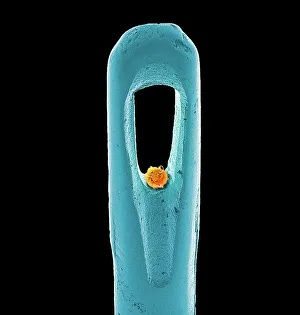
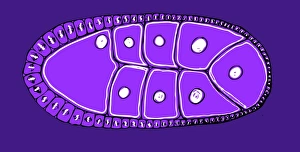
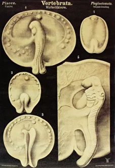
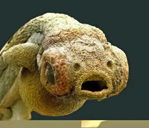
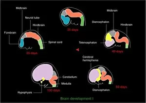
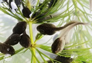
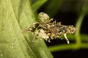
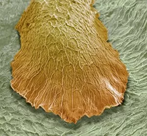
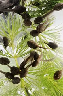
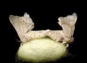
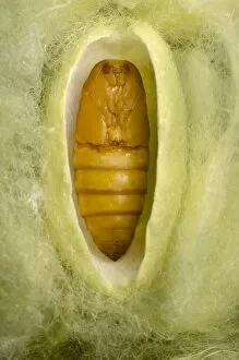
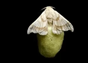
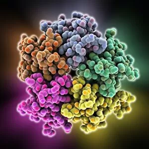
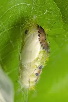
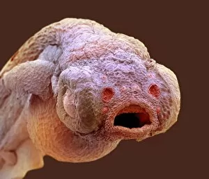
"Unveiling the Wonders of Developmental Biology: From Embryonic Stem Cells to Fetal Brain Development" Embark on a fascinating journey into the realm of developmental biology, where intricate processes shape life itself. In this captivating world, we explore the remarkable transformations that occur from conception to birth and beyond. Delicate as an embryonic stem cell being manipulated by a needle under the watchful eye of scientists, these tiny building blocks hold immense potential for regenerative medicine and understanding human development. Peering through a scanning electron microscope (SEM), we are mesmerized by the intricate head structure of a young newt. Its features reveal nature's precision in crafting complex organisms from simple beginnings. An artwork depicting fish embryo showcases nature's artistic flair in shaping life forms. Each stroke represents the delicate balance between genetic instructions and environmental cues that guide growth and differentiation. In another masterpiece, fetal brain development unfolds before our eyes. Brushstrokes capture the intricacies of neural connections forming within this vital organ, laying down foundations for cognition and consciousness. Observing tadpoles feeding on pond weed reminds us how sustenance fuels growth during early stages of life. These voracious eaters exemplify nature's ingenious mechanisms for ensuring survival through nutrient acquisition. A light micrograph reveals stunning details of a mouse embryo, highlighting its astonishing resemblance to humans at such an early stage. This shared blueprint underscores our evolutionary heritage and offers insights into human development. Witnessing a human fetus nestled comfortably in its mother's womb evokes awe-inspiring wonder at life's miraculous creation. Here lies one of humanity's most profound mysteries - how each individual is shaped with unique traits yet connected by common threads. The bagworm larva captivates with its ability to construct elaborate protective cases around itself using silk and natural materials—an embodiment of adaptation strategies employed throughout evolution to ensure survival amidst changing environments. Traveling back centuries through time, an 1825 artwork showcases the beauty of fetal brain development.