
Diagnosing Collection
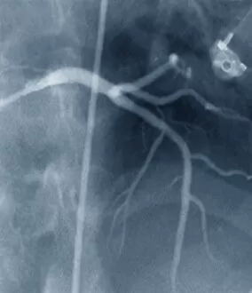
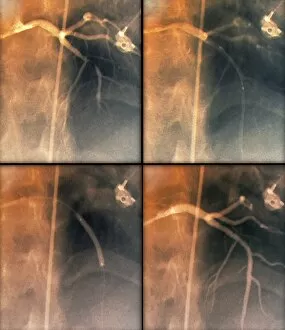
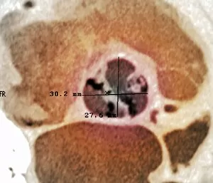
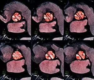
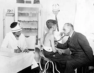
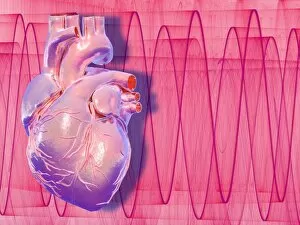
"Unveiling the Art of Diagnosing: From Ancient Practices to Modern Techniques" In this captivating image, a young boy can be seen measuring his temperature


All Professionally Made to Order for Quick Shipping
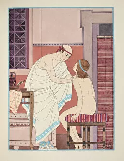
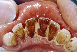
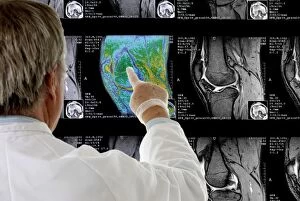
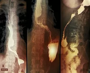
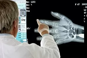
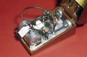
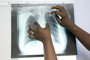
"Unveiling the Art of Diagnosing: From Ancient Practices to Modern Techniques" In this captivating image, a young boy can be seen measuring his temperature, symbolizing the timeless pursuit illnesses. The journey of diagnosis dates back centuries, with pioneers like Abulcasis, an Islamic physician who laid the foundation for medical knowledge. The illustration from "The Works of Hippocrates" showcases an oral examination, highlighting how ancient physicians meticulously observed symptoms and signs to unravel mysteries hidden within the human body. Even then, patients would tremble at the hand of doctors as depicted in another intriguing illustration. Did you know that George Washington himself turned into a physician? This engraving reminds us that even great leaders recognized the importance of understanding ailments and providing care. Richard Morton's contribution cannot be overlooked either; he made significant strides in identifying diseases such as tuberculosis. Dental plaque and tartar are also part of this diagnostic puzzle - their presence revealing more than just dental health but potential systemic issues too. Fast forward to modern times where radiologists examine knee MRI scans with precision (C014 / 1300). X-rays have become invaluable tools in detecting throat cancer (X-rays) while Oriental figures from circa 1830 remind us that different cultures had unique approaches to diagnosis (C017 / 0738). Medical alchemists from the 18th century sought answers through experimentation and exploration. Their practices paved the way for advancements we benefit from today. Lastly, a urine diabetes test (C014 / 1231) serves as a reminder that diagnostics extend beyond visual observations – it involves analyzing bodily fluids too. From ancient wisdom to cutting-edge technology, it has evolved immensely over time, and is through these collective efforts that we continue to unlock medical mysteries and provide better care for all.