Fraud Blocker Skip to main content

Diagnosis Collection (page 27)

"Unlocking the Secrets: The Power of Diagnosis" A seahorse's hidden world revealed through an X-ray, reminding us that diagnosis goes beyond human health

Background imageDiagnosis Collection: Brain examination, conceptual image

Brain examination, conceptual image. Computer artwork of a magnifying glass in front of a brain, representing brain examinations

Background imageDiagnosis Collection: Medical blood analysis

Medical blood analysis. Glass slides being used in a hospital laboratory to analyse a blood sample from a patient, to help diagnose diseases. Photographed at St. Marys Hospital in Lacor, Gulu, Uganda

Background imageDiagnosis Collection: Fractured foot, X-ray

Fractured foot, X-ray
Fractured foot. Coloured X-ray of a foot with a fractured (bright orange area) fourth metatarsal bone. This type of fracture is a common sports injury

Background imageDiagnosis Collection: Skeleton X-ray, artwork

Skeleton X-ray, artwork
Skeleton X-ray, computer artwork. This depiction of a full body X-ray shows the 206 bones of the human skeleton in a normal standing position

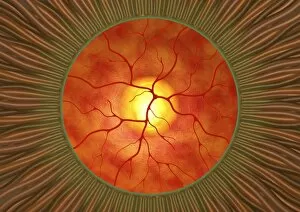
Background imageDiagnosis Collection: Retina in glaucoma, artwork

Retina in glaucoma, artwork
Retina in glaucoma, computer artwork. This view is looking through the pupil (green ring) at the front of the eye, to the retina (orange) at the back of the eye

Background imageDiagnosis Collection: Aortic aneurysm, 3-D CT scan

Aortic aneurysm, 3-D CT scan
3-D Computed Tomography (CT) scan of the abdomen of a patient showing an aortic aneurysm, a bulging of this main artery of the body (red, at centre) which can cause it to rupture if not treated

Background imageDiagnosis Collection: Fractured wrist bone, MRI scan

Fractured wrist bone, MRI scan
Magnetic resonance imaging (MRI) scan (thick slab) of the bones of the wrist, showing a fractured scaphoid bone (almond shaped, centre left)

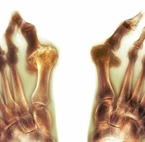
Background imageDiagnosis Collection: X-ray of bunions on the toes

X-ray of bunions on the toes
Coloured X-ray (top view) of the feet of a patient, showing bunions (hallux valgus) on each of the large toes. Bunions are a swelling of the large toe joint which deforms the toes

Background imageDiagnosis Collection: Iliac artery stenosis, 3-D MRI scan

Iliac artery stenosis, 3-D MRI scan
3-D Magnetic resonance imaging (MRI) scan of the abdomen of a patient, showing stenosis (narrowing, centre right) in one of the two iliac arteries which lead into the legs from the abdominal aorta

Background imageDiagnosis Collection: Complete thigh-bone fracture, X-ray

Complete thigh-bone fracture, X-ray
X-ray (back view) of the leg of an adult patient, showing a complete fracture of the thigh bone (femur, running top centre to bottom)

Background imageDiagnosis Collection: Gouty foot, X-ray

Gouty foot, X-ray
Coloured X-ray (top view) of the foot of a patient, showing the big toe affected by gout (gouty arthritis). Gout is a metabolic disease in which uric acid crystals are deposited in joints

Background imageDiagnosis Collection: Bilateral renal artery stenosis, MRA sca

Bilateral renal artery stenosis, MRA sca
Coloured three-dimensional Magnetic Resonance Angiogram (MRA) scan of the abdomen of a male patient aged 77, showing bilateral renal artery stenosis (narrowing)

Background imageDiagnosis Collection: Rheumatoid arthritis of the hands, X-ray

Rheumatoid arthritis of the hands, X-ray
Coloured X-ray (top view) of the hands of a patient, showing rheumatoid arthritis affecting some of the joints at the base of the fingers

Background imageDiagnosis Collection: Splintered arm fracture, X-ray

Splintered arm fracture, X-ray
Coloured X-ray (front view) of the arm of a patient, showing a splintered (multiple) fracture to the upper arm bone (humerus). The bone has broken in three places

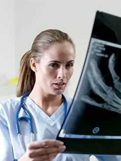
Background imageDiagnosis Collection: Examining X-ray

Examining X-ray
MODEL RELEASED. Examining X-ray. Radiologist examining an X-ray of a patients hand and wrist, looking for broken bones and other disorders

Background imageDiagnosis Collection: Brain tumour, 3-D MRI scan

Brain tumour, 3-D MRI scan
Brain tumour. Coloured three-dimensional model of a male head, made up from numerous magnetic resonance imaging (MRI) scans

Background imageDiagnosis Collection: Heart and lungs, 3D CT scan

Heart and lungs, 3D CT scan
Heart and lungs, 3D-CT scan. Coloured 3-D computed tomography (CT) scan of the heart (middle centre) and lungs, showing the small airways (bronchioles) of the lungs. The rib cage surrounds the lungs

Background imageDiagnosis Collection: Patient has Hand X-Rayed

Patient has Hand X-Rayed
The French engineer, Seguy, x- rays a patients hand

Background imageDiagnosis Collection: CALANDER SAYS NO

CALANDER SAYS NO
The CALANDER (a kind of lark) knows if sick people will recover; held over the sickbed, if it turns towards the patient s/he will recover, if not (as here) s/he won t

Background imageDiagnosis Collection: Doctor discussing medical records

Doctor discussing medical records
MODEL RELEASED. Medical records on a doctors desk. The doctor is using a telephone and looking at one of the records

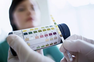
Background imageDiagnosis Collection: Urine analysis

Urine analysis
MODEL RELEASED. Urine analysis. Generalpractice doctor comparing a multiple test stick(Multistix) to its reference chart. The test stickhas been placed into a sample of a patients urineto test for a

Background imageDiagnosis Collection: Throat examination

Throat examination
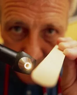
MODEL RELEASED. Throat examination. Generalpractice (GP) doctor holding a tongue depressor(right) and a viewing instrument (left)

Background imageDiagnosis Collection: Examining the Ear

Examining the Ear
Examining the ear

Background imageDiagnosis Collection: X-Ray Tube / Vickers / 1936

X-Ray Tube / Vickers / 1936
Million volt X-ray tube; developed by Metropolitan Vickers; installed in St. Bartholomews Hospital

Background imageDiagnosis Collection: Dr Diocles X-Ray / 1926

Dr Diocles X-Ray / 1926
Dr Diocles radio stereoscope presents 3-D image of the subject

Background imageDiagnosis Collection: Diagnosis from Urine

Diagnosis from Urine
Urine diagnosis chart


All Professionally Made to Order for Quick Shipping

"Unlocking the Secrets: The Power of Diagnosis" A seahorse's hidden world revealed through an X-ray, reminding us that diagnosis goes beyond human health. Unveiling the spectrum: a color blindness test helping us understand how our eyes perceive the world differently. Eyesight test chart guiding us towards clarity and precision in diagnosing visual impairments. Peering into fractures: a broken wrist bone captured on X-ray, aiding doctors in accurate diagnosis and treatment plans. Exploring beneath the surface: a panoramic dental X-ray exposing hidden oral issues for comprehensive diagnoses. Empowering individuals with knowledge: home cholesterol test kits providing early detection for proactive health management. Saving lives through swift intervention: Pneumothorax treatment guided by an X-ray to restore lung function effectively. Knees tell stories too: a normal knee showcased on an X-ray, enabling precise diagnoses of joint conditions and injuries. Delving deeper into mysteries within: full body scans via MRI revealing intricate details for accurate medical assessments. Artistry meets science as ECGs portray the rhythm of a healthy heart, serving as diagnostic masterpieces in healthcare settings. Battling tuberculosis head-on with an X-ray, unveiling its presence so prompt treatments can be initiated promptly