
Diagnostic Collection
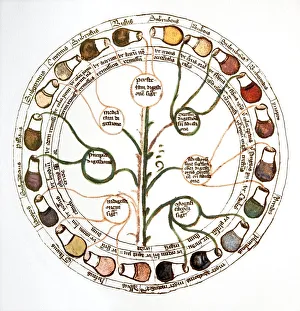
"Unlocking the Secrets of Health: Exploring the World Tools" Medieval Urine Wheel: From ancient times to modern medicine


All Professionally Made to Order for Quick Shipping
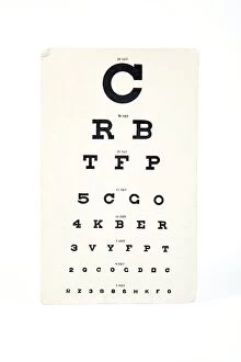
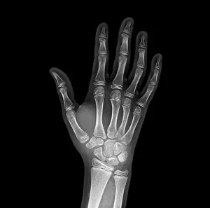
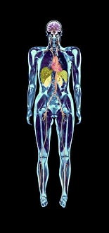
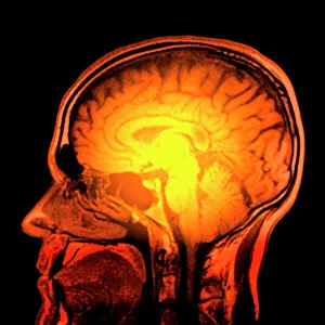
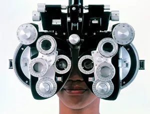
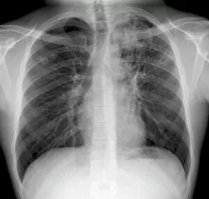
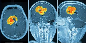
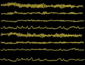
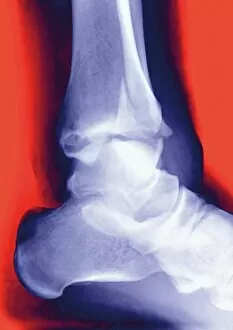
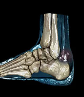
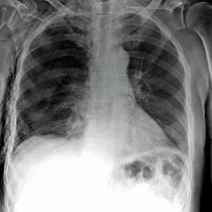
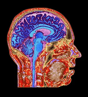
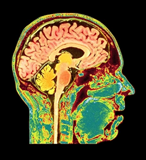
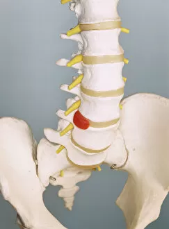
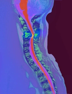
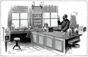
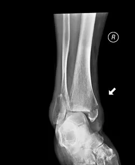
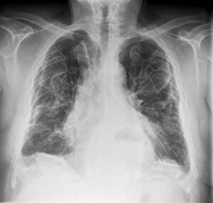
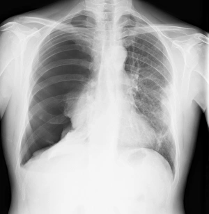
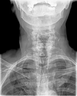
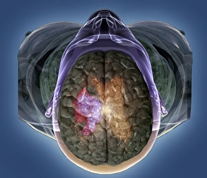
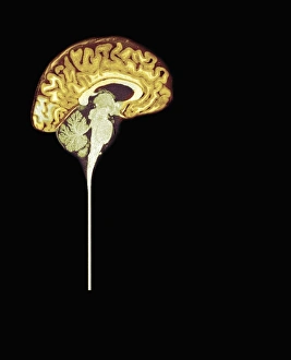
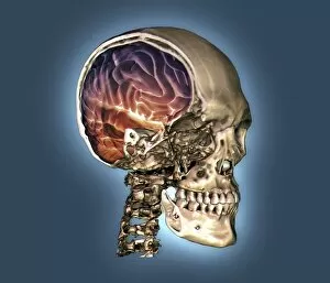
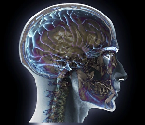
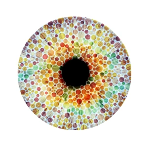
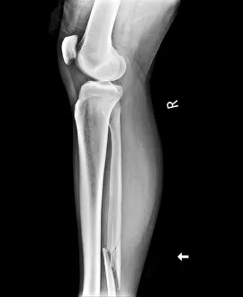
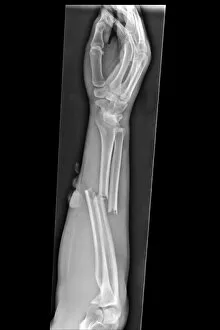
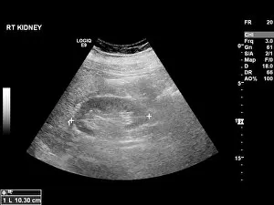
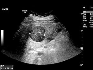
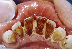
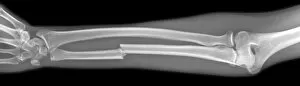
"Unlocking the Secrets of Health: Exploring the World Tools" Medieval Urine Wheel: From ancient times to modern medicine, urine analysis has been a diagnostic tool used to detect various health conditions. Eyesight Test Chart: A simple yet crucial diagnostic tool that helps determine visual acuity and identify potential eye problems. Broken Wrist Bone, X-ray C017 / 7187: X-rays provide a clear view of fractured bones, aiding in accurate diagnosis and guiding treatment plans. Panoramic Dental X-ray: This comprehensive imaging technique reveals dental issues like cavities, impacted teeth, or jaw abnormalities for effective treatment planning. Home Cholesterol Test Kit: Empowering individuals with easy-to-use kits to monitor cholesterol levels at home and take proactive steps towards heart health. Full Body Scan, MRI Scan: Non-invasive and highly detailed imaging technology that allows doctors to visualize internal organs and diagnose complex medical conditions accurately. Brain Anatomy, MRI Scan: Advanced brain imaging enables neurologists to study intricate structures within the brain for precise diagnoses of neurological disorders. ECGs of a Normal Heart Rate, Artwork: Electrocardiograms help assess heart function by recording electrical activity; this artwork showcases what a healthy heartbeat looks like on paper. Tuberculosis, X-ray: Chest x-rays play a vital role in diagnosing tuberculosis by detecting characteristic lung abnormalities associated with this infectious disease. Normal EEG Readout of the Brain's Alpha Waves: Electroencephalography records brain wave patterns helping neurologists evaluate brain function and diagnose epilepsy or sleep disorders effectively. Eye Examination:A thorough examination assessing vision sharpness (visual acuity), peripheral vision tests (visual field), color perception evaluation among others is essential for maintaining good eye health Fractured Ankle, X-Ray.