
Digestive Enzyme Collection
Caption: Unveiling the Intricate World of Digestive Enzymes In this captivating light micrograph, we delve into the fascinating realm of digestive enzymes


All Professionally Made to Order for Quick Shipping
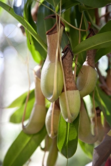
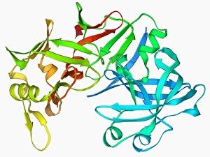
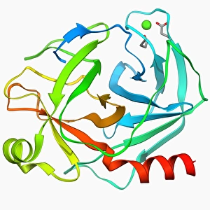
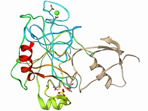
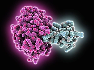
Caption: Unveiling the Intricate World of Digestive Enzymes In this captivating light micrograph, we delve into the fascinating realm of digestive enzymes. The Islet of Langerhans takes center stage, showcasing its vital role in our body's intricate digestion process. As we explore further, we encounter a remarkable Nepenthes - a tropical pitcher plant known for its unique ability to produce digestive enzymes. Just like these carnivorous plants, our bodies possess an array of specialized enzymes that aid in breaking down complex molecules. Zooming in closer, we witness the power of pepsin - an enzyme found in our stomachs. Its molecular structure (F006 / 9767) reveals its efficiency in breaking down proteins into smaller fragments for better absorption and utilization by our bodies. Next on display is pepsinogen (F006 / 9710), the precursor molecule to pepsin. This inactive form safeguards us from self-digestion until it reaches the acidic environment within our stomachs where it transforms into active pepsin. Our exploration continues with trypsin (F006 / 9634), another crucial enzyme responsible for protein digestion. We observe its dynamic structure along with inhibitors (F006 / 9633) that regulate its activity, preventing excessive breakdown and maintaining balance within our digestive system. Further unveiling trypsinogen (F006 / 9517), we discover how this dormant molecule awaits activation before transforming into trypsin. Such meticulous regulation ensures optimal enzymatic function while safeguarding against unwanted premature activation. Delving deeper still, we encounter additional trypsin molecules alongside inhibitors (C015 / 8436). These inhibitors play a pivotal role in fine-tuning enzymatic activities to prevent any disruption or imbalance within the delicate ecosystem of digestion. Returning once again to marvel at the Islet of Langerhans through multiple light micrographs, we appreciate their significance in regulating blood sugar levels.