
Digestive System Collection (page 3)
The digestive system is a complex network of organs and processes that allows our bodies to break down food and absorb nutrients


All Professionally Made to Order for Quick Shipping
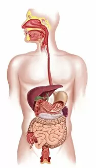
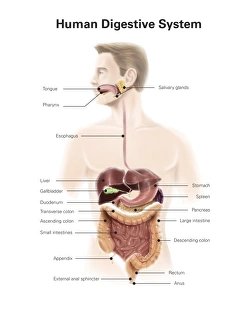
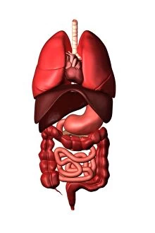
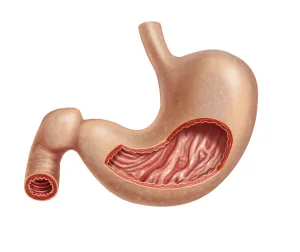
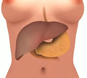
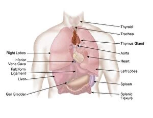
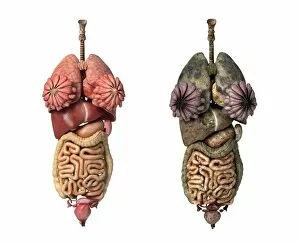
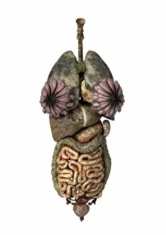
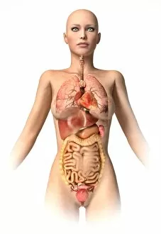
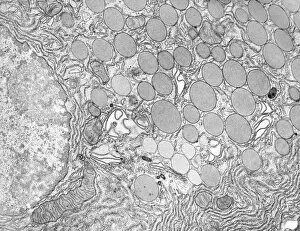
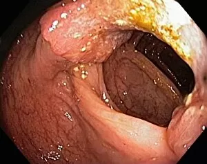
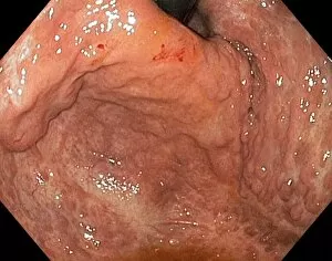
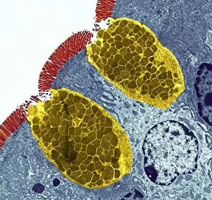
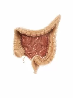
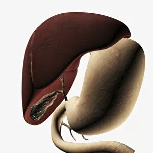
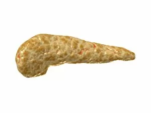
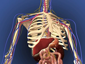
The digestive system is a complex network of organs and processes that allows our bodies to break down food and absorb nutrients. From the gastrointestinal nematodes that inhabit our intestines to the medical illustration of an appendix with appendicitis, every component plays a vital role in this intricate system. Bee anatomy, depicted through artwork, showcases how these tiny creatures have their own unique digestive systems. Meanwhile, a scanning electron microscope (SEM) image reveals the Salmonella bacteria responsible for causing foodborne illnesses. Another SEM image focuses on the small intestine's surface, highlighting its crucial function in nutrient absorption. Similarly, we see the gallbladder's surface under SEM examination, emphasizing its role in storing bile produced by the liver. C. Elegans worms are also featured here as they provide valuable insights into understanding human digestion at a microscopic level. A light micrograph captures their intricate structure and behavior within this fascinating ecosystem. Moving beyond microscopic organisms, we encounter a muskrat sitting on the shore of a pond indulging in grass consumption—an example of herbivorous digestion within nature's cycle. An X-ray image provides us with an inside look at an inflamed appendix—a condition known as appendicitis—underscoring both its importance and potential health risks if left untreated. Lastly, artwork showcasing pancreas anatomy reminds us of this organ's significant contribution to digestion through hormone production such as insulin regulation.