Fraud Blocker Skip to main content

Dissected Collection (page 2)

"Unveiling the Intricacies of Life: A Journey through Dissected Hints" Step into the world of Leonardo da Vinci's skull anatomy

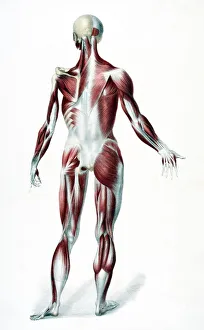
Background imageDissected Collection: Back Of The Male Human Body Showing Muscles Sinews And Bones From The Vessels Of The Human Body

Back Of The Male Human Body Showing Muscles Sinews And Bones From The Vessels Of The Human Body Edited By Jones Quain And William Wilson Published London By Taylor And Watson 1837

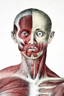
Background imageDissected Collection: Head And Shoulders Of The Male Human Body Showing Muscles Sinews And Bones From The Vessels Of The

Head And Shoulders Of The Male Human Body Showing Muscles Sinews And Bones From The Vessels Of The Human Body Edited By Jones Quain And William Wilson Published London By Taylor And Watson 1837

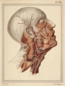
Background imageDissected Collection: Face and neck nerves, 1825 artwork

Face and neck nerves, 1825 artwork
Face and neck nerves. Head and neck dissected to show the superficial nerves (white). The parotid gland (normally present below the ear)
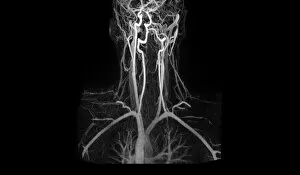
Background imageDissected Collection: Artery damage, MRA scan

Artery damage, MRA scan
Artery damage. Magnetic resonance angiography (MRA) scan of the neck of a 23-year-old male motorcycle crash victim. Dissection of the right internal carotid artery (left)

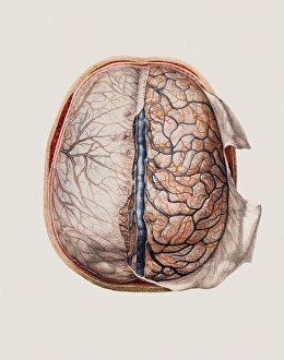
Background imageDissected Collection: Brain meninges

Brain meninges, historical anatomical artwork. This cranial (top) view of the brain (with front at the top) and its surrounding protective meninges, which have been partially dissected away

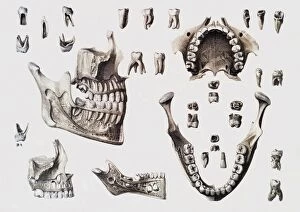
Background imageDissected Collection: Dental anatomy

Dental anatomy. Historical anatomical artwork of healthy and diseased human teeth and jaws. The teeth and jaws are seen from several different directions

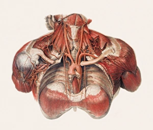
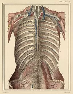
Background imageDissected Collection: Blood vessels of chest and neck

Blood vessels of chest and neck
Blood vessels of the chest and neck, historical anatomical artwork. The thoracic (chest) cavity has been dissected in this anterior (front) view to reveal part of the aorta

Background imageDissected Collection: Aortic dissection, 3D CT scan

Aortic dissection, 3D CT scan
Aortic dissection. 3D computed tomography (CT) scan of a 67-year-old man with an aortic dissection. The aorta, the bodys main artery, is running vertically down centre (red)

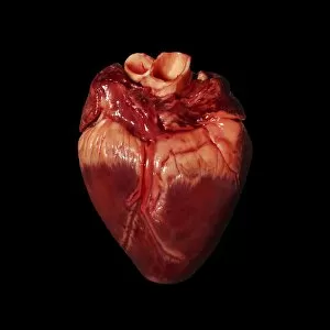
Background imageDissected Collection: Pigs heart

Pigs heart. The heart of a pig is anatomically similar to that of a human. The heart is a hollow muscular organ that pumps blood around the body

Background imageDissected Collection: Skeleton of a pigeon and a collection of pigeon feathers

Skeleton of a pigeon and a collection of pigeon feathers

Background imageDissected Collection: Cross section of a heart

Cross section of a heart
Cross section of a human heart showing the various chambers and valves. Date: 1910

Background imageDissected Collection: Cross-section through skeleton of Radiated tortoise (Astrochelys radiata), side view

Cross-section through skeleton of Radiated tortoise (Astrochelys radiata), side view

Background imageDissected Collection: Separated parts of human skull

Separated parts of human skull

Background imageDissected Collection: Twisted-pair cabling, side view

Twisted-pair cabling, side view

Background imageDissected Collection: Internal anatomy of frog, dissected with its internal organs hanging out of its belly

Internal anatomy of frog, dissected with its internal organs hanging out of its belly

Background imageDissected Collection: Segmented exoskeleton of a beetle separated into parts including the head with large mandibles

Segmented exoskeleton of a beetle separated into parts including the head with large mandibles, wings and carapace and legs

Background imageDissected Collection: Parts of a crayfish

Parts of a crayfish

Background imageDissected Collection: Thoracic aorta, 1825 artwork

Thoracic aorta, 1825 artwork
Thoracic aorta. Dissection of the chest cavity with the heart and lungs and other organs removed, showing the thoracic portion of the aorta (red), the main body artery

Background imageDissected Collection: Intestinal veins, 1825 artwork

Intestinal veins, 1825 artwork
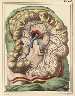
Intestinal veins. Dissection of the abdominal cavity showing the inferior mesenteric vein (blue) that drains blood from the large intestine

Background imageDissected Collection: Diaphragm anatomy, 1831 artwork

Diaphragm anatomy, 1831 artwork
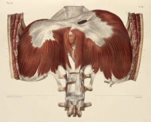
Diaphragm anatomy, posterior view. The diaphragm is the white area at top, connected to surrounding muscles (red). Part of the spine is at bottom, and ribs at left and right

Background imageDissected Collection: Liver and stomach nerves, 1825 artwork

Liver and stomach nerves, 1825 artwork
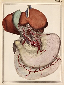
Liver and stomach nerves. Extensive network of nerves (white) supplying the liver (upper left) and stomach (lower right). These nerves networks (plexuses)

Background imageDissected Collection: Intestinal arteries, 1825 artwork

Intestinal arteries, 1825 artwork
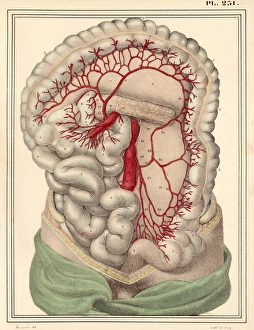
Intestinal arteries. Dissection of the abdomen, showing the inferior mesenteric artery (red) and its branches that supply oxygenated blood to the large intestine

Background imageDissected Collection: Lower leg and foot fascia, 1831 artwork

Lower leg and foot fascia, 1831 artwork
Lower leg and foot fascia. Lateral views of the partitions of the fascia of the muscles of the leg and foot. Fascia are fibrous connective tissues that surround muscles and other tissues

Background imageDissected Collection: Chest lymph vessels, 1825 artwork

Chest lymph vessels, 1825 artwork
Chest lymph vessels. Dissection of the thoracic cavity showing lymphatic vessels (white) and nodes. The central chest lymph vessel is the thoracic canal, running parallel to the backbone (centre)

Background imageDissected Collection: Heart-lung anatomy, 1825 artwork

Heart-lung anatomy, 1825 artwork
Heart-lung anatomy. Axial section through the chest at the level of the fifth thoracic vertebra (T5). The front of the body is at top

Background imageDissected Collection: Abdominal aorta, 1825 artwork

Abdominal aorta, 1825 artwork
Abdominal aorta. Dissection, with some organs removed, showing the abdominal portion of the aorta (red), the main body artery

Background imageDissected Collection: Brain membrane vessels, 1825 artwork

Brain membrane vessels, 1825 artwork
Brain membrane vessels. Axial section through the skull showing the blood vessels (red) of two of the brain membranes, the arachnoid mater and pia mater

Background imageDissected Collection: Face and neck muscles, 1831 artwork

Face and neck muscles, 1831 artwork
Face and neck muscle. Anterior view of the deep layers of muscles of the face and neck, including the eyes and mouth. Insets show details of the lips, chin and eyes

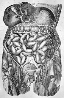
Background imageDissected Collection: Abdominal organs, 1880 artwork C017 / 6912

Abdominal organs, 1880 artwork C017 / 6912
Abdominal organs. Historical anatomical artwork of a dissected abdomen seen from the front, showing the internal organs. These include the intestines, bladder, stomach and liver

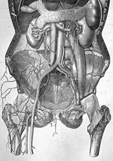
Background imageDissected Collection: Deep abdominal organs, 1880 artwork C017 / 6913

Deep abdominal organs, 1880 artwork C017 / 6913
Deep abdominal organs. Historical anatomical artwork of a dissected abdomen seen from the front, showing the deep-seated organs and major blood vessels after the removal of the intestines

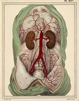
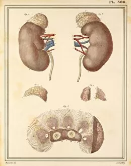
Background imageDissected Collection: Kidney anatomy, 1825 artwork

Kidney anatomy, 1825 artwork
Kidney anatomy. At top are the left and right kidneys (brown) with arteries (red), veins (blue), ureters (white) and adrenal glands (cream, on top of kidneys)

Background imageDissected Collection: Neck muscle anatomy, 1831 artwork

Neck muscle anatomy, 1831 artwork
Neck muscle anatomy. Various views of the anatomy of the muscles of the neck. The main artwork shows the neck seen from the front

Background imageDissected Collection: Atlanto-occipital joint, artwork C016 / 6554

Atlanto-occipital joint, artwork C016 / 6554
Atlanto-occipital joint. Artwork of the bones (yellow) and ligaments (blue) of the atlanto-occipital joint, as seen from behind (posterior view)

Background imageDissected Collection: Liver anatomy, artwork C016 / 7001

Liver anatomy, artwork C016 / 7001
Liver anatomy. Artwork of a frontal view of the liver, dissected to show some of its internal anatomy. The liver, subdivided into lobes, is the largest gland in the human body

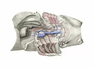
Background imageDissected Collection: Shark heart-gill anatomy, artwork C016 / 6847

Shark heart-gill anatomy, artwork C016 / 6847
Shark heart-gill anatomy. Artwork of the heart and gill anatomy of a shark, seen from the underside, with the head at right (teeth at centre right)

Background imageDissected Collection: Atlanto-occipital joint, artwork C016 / 6555

Atlanto-occipital joint, artwork C016 / 6555
Atlanto-occipital joint. Artwork of the bones (yellow) and ligaments (blue) of the atlanto-occipital joint, as seen from behind (posterior view)

Background imageDissected Collection: Dissected mouse, illustration C018 / 0781

Dissected mouse, illustration C018 / 0781
Dissected mouse (Mus musculus), illustration

Background imageDissected Collection: Sectional dissected head, 19th century C017 / 0705

Sectional dissected head, 19th century C017 / 0705
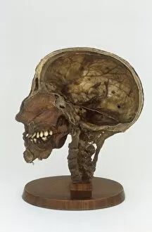
Dissected cross section of a human head and neck with the brain removed. Anatomy of the skull, spine, teeth and sinuses can be seen. This artefact dates from the nineteenth century

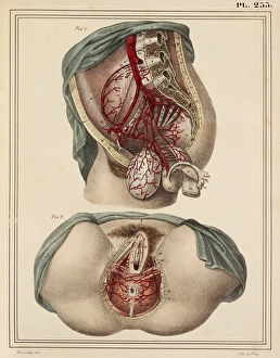
Background imageDissected Collection: Female groin arteries, 1825 artwork

Female groin arteries, 1825 artwork
Female groin arteries. Dissections of a female groin to show areas supplied by the internal iliac artery (red) and its branches

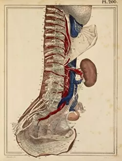
Background imageDissected Collection: Lumbar spinal nerves, 1825 artwork

Lumbar spinal nerves, 1825 artwork
Lumbar spinal nerves. Lateral view of the lower vertebral column, showing nerves (white) of the sympathetic nervous system. The pelvic ilium bone has been removed

Background imageDissected Collection: Trunk arteries, 1825 artwork

Trunk arteries, 1825 artwork
Trunk arteries. Dissection showing the internal mammary arteries (red, in chest) and the epigastric arteries (red, in groin and abdomen), from a mans trunk (torso)

Background imageDissected Collection: Lower leg arteries, 1825 artwork

Lower leg arteries, 1825 artwork
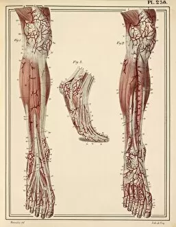
Lower leg arteries. Anterior (frontal) view of the arteries (red) and muscles and ligaments of the lower leg and foot. At left are the superficial arteries

Background imageDissected Collection: Neck and shoulder arteries, 1825 artwork

Neck and shoulder arteries, 1825 artwork
Neck and shoulder arteries (red). Dissection showing the right carotid arteries of the neck, and the right subclavian artery of the shoulder

Background imageDissected Collection: Lung lymph vessels, 1825 artwork

Lung lymph vessels, 1825 artwork
Lung lymph vessels. Dissected right lung showing its lymph vessels (white). The dark bands are fissures between the different lobes of the lung

Background imageDissected Collection: Pulmonary nerve plexus, 1825 artwork

Pulmonary nerve plexus, 1825 artwork
Pulmonary nerve plexus. View from behind, with the thoracic spine removed, of the posterior pulmonary plexus, a confluence of nerves (white)

Background imageDissected Collection: Liver and stomach arteries, 1825 artwork

Liver and stomach arteries, 1825 artwork
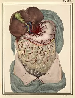
Liver and stomach arteries. Dissection of the abdomen, showing the arteries (red) of the liver (brown, upper left), stomach (pink, upper centre), and the intestines (grey with yellow fat)

Background imageDissected Collection: Throat muscles, 1831 artwork

Throat muscles, 1831 artwork
Throat muscles. Posterior view of the muscles of the pharynx (part of the throat). The pharynx is at centre, with the base of the head at top and the upper part of the chest cavity at bottom

Background imageDissected Collection: Veins of the stomach, 1825 artwork

Veins of the stomach, 1825 artwork
Veins of the stomach. Abdominal dissection showing the veins (blue) of the stomach (centre) and the greater omentum (lower centre, yellow)


All Professionally Made to Order for Quick Shipping

"Unveiling the Intricacies of Life: A Journey through Dissected Hints" Step into the world of Leonardo da Vinci's skull anatomy, where every curve and crevice holds secrets to our existence. Marvel at the intricate network of blood vessels that nourish our brain, as depicted in his mesmerizing artwork. The heart, a symbol of life itself, beats tirelessly within us. Explore its chambers and valves, understanding its vital role in sustaining our being. Witness the delicate dance between arteries and veins that carry life-giving oxygen throughout our bodies. Delve into history with an 1825 artwork showcasing male groin arteries - a testament to early anatomical studies. Discover how these hidden pathways contribute to our overall health and well-being. Venture further into the realm of dissection as we unravel the complex relationship between the heart and lungs. Witness their interdependence, understanding how they work harmoniously to ensure proper oxygenation for survival. Behold an 1825 artwork depicting head and chest arteries; a visual representation of scientific exploration from centuries past. Marvel at how these intricate networks sustain life by delivering essential nutrients throughout our bodies. Explore beyond mere surface appearances as we confront liver cirrhosis – a stark reminder of what can happen when this vital organ is compromised. Gain insight into medical conditions that challenge us while appreciating the resilience of human physiology. Witness scientists meticulously working with ragworm specimens – emblematic of tireless dedication towards unlocking nature's mysteries. Celebrate their unwavering commitment to advancing knowledge for generations to come. Embark on a journey through facial nerves, discovering their role in conveying emotions across countless expressions etched upon our faces. Appreciate how these invisible threads connect mind and body in profound ways. Transport yourself back in time to witness Eustachian tube syringing practices from the 18th century – an intriguing glimpse into historical medical techniques aimed at restoring balance within fragile auditory systems.