
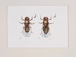
Drosophila Melanogaster Collection
"Drosophila melanogaster: A Fascinating World Under the SEM" Step into the microscopic world of Drosophila melanogaster, commonly known as the fruit fly


All Professionally Made to Order for Quick Shipping
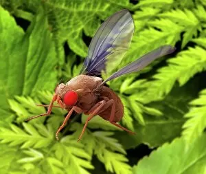
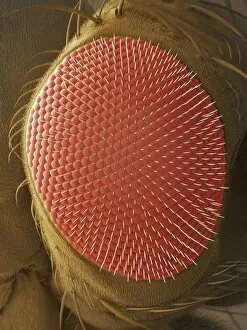
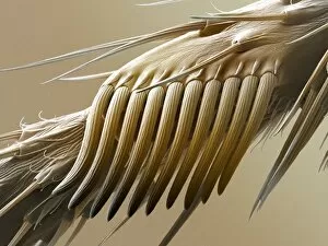
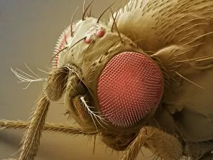
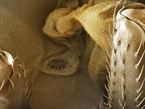
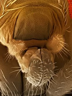
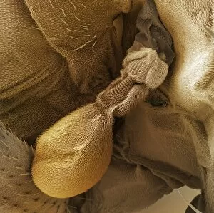
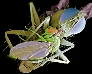
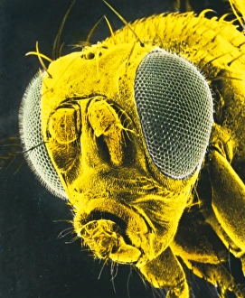
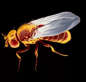
"Drosophila melanogaster: A Fascinating World Under the SEM" Step into the microscopic world of Drosophila melanogaster, commonly known as the fruit fly. Despite its small size, this tiny insect holds immense significance in scientific research. With the help of scanning electron microscopy (SEM), we can explore various intricate features that make these flies truly remarkable. The fruit fly brain, illustrated in C018 / 0791, showcases a complex network of neurons responsible for their behavior and sensory perception. Moving on to their compound eye, SEM reveals a mesmerizing array of facets that enable them to perceive motion and color with incredible precision. Intriguingly, male fruit flies possess a unique structure called the sex comb which aids in courtship rituals. SEM imaging unveils its fine bristles arranged meticulously for attracting potential mates. The antenna is another captivating feature; SEM uncovers its delicate sensory structures that allow these insects to detect pheromones and navigate their environment effectively. Examining further under SEM, we discover astonishing details about the fruit fly's head - from its intricate mouthpart or proboscis used for feeding (SEM) to specialized balance organs aiding flight control (SEM). Even their minuscule feet reveal fascinating adaptations when observed closely through an SEM lens. Venturing deeper into their anatomy, we encounter spiracles – tiny openings along their body surface allowing gas exchange – captured beautifully by SEM imagery. Additionally, illustrations like C018 / 0784 showcase embryonic stages of these flies' development providing valuable insights into genetics and developmental biology. Finally, let us not forget about those iconic wings. Illustrated in C018 / 0792 they can essential for flight and exhibit stunning patterns unique to each individual fly. Drosophila melanogaster may be diminutive but exploring it through advanced techniques like SEM unravels a hidden world full of wonders waiting to be discovered.