Fraud Blocker Skip to main content

Electrocardiography Collection

Electrocardiography, commonly known as ECG, is a vital tool in the field of cardiology

Background imageElectrocardiography Collection: ECGs of a normal heart rate, artwork

ECGs of a normal heart rate, artwork
Electrocardiograms (ECG) of a normal heart rate, artwork. An ECG measures the electrical activity of the heart. The wave on the ECG appears upwards or downwards depending upon whether the electrical

Background imageElectrocardiography Collection: Irregular heartbeat

Irregular heartbeat. Conceptual image of a chest X-ray overlaid with an electrocardiogram (heart monitor) trace showing extrasystole

Background imageElectrocardiography Collection: ECG, artwork F006 / 4606

ECG, artwork F006 / 4606
ECG. Computer artwork of an electrocardiogram (ECG) showing a normal heart rate

Background imageElectrocardiography Collection: ECG, computer artwork

ECG, computer artwork
ECG. Computer artwork of an electrocardiogram (ECG) showing a normal heart rate

Background imageElectrocardiography Collection: Extrasystole heartbeats, ECG artwork

Extrasystole heartbeats, ECG artwork. Diagram showing the electrical activity of the atrium (top) and ventricle (bottom) of a human heart during extrasystole

Background imageElectrocardiography Collection: Cardiology, conceptual image C017 / 7718

Cardiology, conceptual image C017 / 7718
MODEL RELEASED. Cardiology, conceptual image

Background imageElectrocardiography Collection: Cardiac patient in an ambulance C016 / 7443

Cardiac patient in an ambulance C016 / 7443
Cardiac patient in an ambulance. Paramedic preparing to perform an electrocardiogram (ECG) on a 50-year-old cardiac patient inside an ambulance

Background imageElectrocardiography Collection: ECG trace C017 / 7744

ECG trace C017 / 7744
Electrocardiogram (ECG) trace. An ECG measures the electrical activity of the heart

Background imageElectrocardiography Collection: ECG trace C017 / 7743

ECG trace C017 / 7743
Electrocardiogram (ECG) trace. An ECG measures the electrical activity of the heart

Background imageElectrocardiography Collection: Cardiology, conceptual image C017 / 7719

Cardiology, conceptual image C017 / 7719
MODEL RELEASED. Cardiology, conceptual image

Background imageElectrocardiography Collection: Cardiology, conceptual image C017 / 7717

Cardiology, conceptual image C017 / 7717
MODEL RELEASED. Cardiology, conceptual image

Background imageElectrocardiography Collection: Obesity clinic C013 / 7681

Obesity clinic C013 / 7681
MODEL RELEASED. Obesity clinic. Male patient undergoing an electrocardiography (ECG) test

Background imageElectrocardiography Collection: Obesity clinic C013 / 7663

Obesity clinic C013 / 7663
MODEL RELEASED. Obesity clinic. Male patient undergoing an electrocardiography (ECG) test

Background imageElectrocardiography Collection: Obesity clinic C013 / 7661

Obesity clinic C013 / 7661
MODEL RELEASED. Obesity clinic. Male patient undergoing an electrocardiography (ECG) test

Background imageElectrocardiography Collection: Obesity clinic C013 / 7664

Obesity clinic C013 / 7664
MODEL RELEASED. Obesity clinic. Male patient undergoing an electrocardiography (ECG) test

Background imageElectrocardiography Collection: Obesity clinic C013 / 7516

Obesity clinic C013 / 7516
MODEL RELEASED. Obesity clinic. Male patient undergoing an electrocardiography (ECG) test

Background imageElectrocardiography Collection: Obesity clinic C013 / 7470

Obesity clinic C013 / 7470
MODEL RELEASED. Obesity clinic. Male patient undergoing an electrocardiography (ECG) test

Background imageElectrocardiography Collection: ECG electrode

ECG electrode on a patient. Electrodes respond to electrical impulses in the heart. They are stuck to the skin with a removable sticky pad (white)

Background imageElectrocardiography Collection: ECG

ECG. Computer artwork of an electrocardiogram (ECG) showing a normal heart rate. An ECG measures the electrical activity of the heart

Background imageElectrocardiography Collection: Atrial fibrillation, artwork

Atrial fibrillation, artwork
Atrial fibrillation. Artwork of a section through a human heart during atrial fibrillation, a rapid, irregular heartbeat. Two electrocardiogram traces (ECGs) are seen at centre left and upper right

Background imageElectrocardiography Collection: Artwork of healthy ECG traces of the heart

Artwork of healthy ECG traces of the heart
Healthy ECG heart traces. Computer-enhanced image of many electrocardiogram (ECG) traces of the normal heartbeat. These ECG heart traces show a healthy heart rhythm

Background imageElectrocardiography Collection: Artwork of sectioned heart with healthy ECG trace

Artwork of sectioned heart with healthy ECG trace
Heart nerve conduction. Illustration of a section through a human heart with an electrocardiogram (ECG) trace. The heart is a hollow muscle that pumps blood around the body

Background imageElectrocardiography Collection: Computer artwork of healthy ECG trace of the heart

Computer artwork of healthy ECG trace of the heart
Healthy ECG heart trace. Computer-enhanced image of an electrocardiogram (ECG) trace of the normal heartbeat. This ECG heart trace shows a healthy heart rhythm

Background imageElectrocardiography Collection: Computer graphic of ECG trace on a healthy heart

Computer graphic of ECG trace on a healthy heart

Background imageElectrocardiography Collection: ECG and man running

ECG and man running. Computer artwork of an electrocardiogram (ECG) showing a normal heart rate and the outline of a man running. An ECG measures the electrical activity of the heart

Background imageElectrocardiography Collection: ECG of a normal heart rate

ECG of a normal heart rate
Electrocardiogram. Hands holding an electrocardiogram (ECG) of a normal heart rate. An ECG measures the electrical activity of the heart

Background imageElectrocardiography Collection: Doctor puts electrodes on mans chest for ECG test

Doctor puts electrodes on mans chest for ECG test

Background imageElectrocardiography Collection: Doctor examines a male patients ECG trace

Doctor examines a male patients ECG trace
MODEL RELEASED. ECG examination. Doctor examines a patients electrocardiograph (ECG) trace taken in order to monitor the health of his heart

Background imageElectrocardiography Collection: Doctor carries out an ECG examination on a man

Doctor carries out an ECG examination on a man
MODEL RELEASED. ECG examination. Doctor carries out an electrocardiograph (ECG) examination on a man to monitor the health of his heart

Background imageElectrocardiography Collection: Healthy electrocardiograph and vitalograph traces

Healthy electrocardiograph and vitalograph traces

Background imageElectrocardiography Collection: Atrial fibrillation

Atrial fibrillation. Artwork of a section through a human heart during atrial fibrillation, a rapid, irregular heartbeat. In the background are ECG (electrocardiogram) traces

Background imageElectrocardiography Collection: ECG test

ECG test
Model Released. Ecg Test

Background imageElectrocardiography Collection: Heart fitness test

Heart fitness test
MODEL RELEASED. Heart fitness test

Background imageElectrocardiography Collection: ECG and brain outline

ECG and brain outline
ECG and brain. Computer artwork of an electrocardiogram (ECG) showing a normal heart rate and the outline of a human brain. An ECG measures the electrical activity of the heart

Background imageElectrocardiography Collection: Heart monitoring

Heart monitoring
MODEL RELEASED. Heart monitoring. Close-up of electrodes on a patients chest during an electrocardiography (ECG) examination. The electrodes are used to measure the electrical activity of his heart

Background imageElectrocardiography Collection: Mareys heart machines, 19th century

Mareys heart machines, 19th century
Mareys heart machines. 19th-century artworks of the devices developed in 1881 by the French scientist Etienne-Jules Marey (1830-1904) for the study of the circulatory system

Background imageElectrocardiography Collection: Placing of electrodes for ECG, artwork

Placing of electrodes for ECG, artwork
Placing of electrodes for electrocardiogram (ECG), artwork. This diagram shows where the six electrodes attached to the chest should be placed. One electrode is also placed on each arm and leg

Background imageElectrocardiography Collection: ECG of a normal heart rate, artwork

ECG of a normal heart rate, artwork
Electrocardiogram (ECG) of a normal heart rate, artwork. An ECG measures the electrical activity of the heart. Contractions are caused by electrical signals between the upper (atrial)

Background imageElectrocardiography Collection: Electrical conduction of the heart

Electrical conduction of the heart. Artwork showing the impulse conduction system (yellow) of the human heart (left) and an electrocardiogram (ECG) of a normal heart rate (right)

Background imageElectrocardiography Collection: ECG electrode placement, artwork

ECG electrode placement, artwork
ECG electrode placement. Computer artwork showing the location of the 10 electrodes needed to perform a 12 lead electrocardiogram (ECG) test

Background imageElectrocardiography Collection: Heartbeat, conceptual image

Heartbeat, conceptual image
heartbeat

Background imageElectrocardiography Collection: ECG, conceptual artwork

ECG, conceptual artwork
ECG. Computer artwork of a spiral electrocardiogram (ECG) showing a normal heart rate


All Professionally Made to Order for Quick Shipping

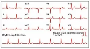
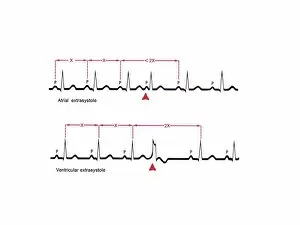
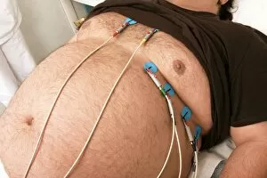
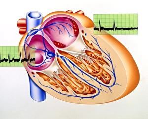
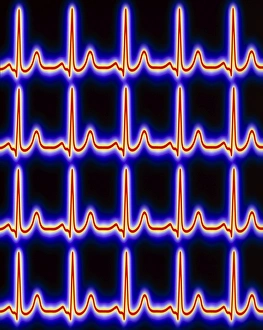
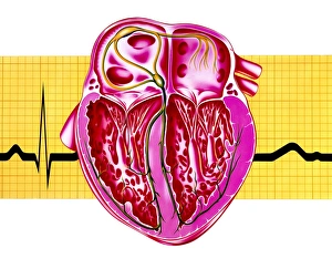
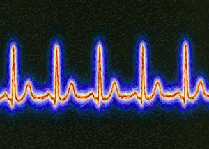
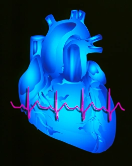
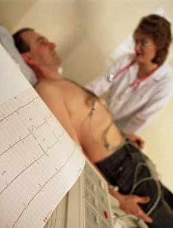
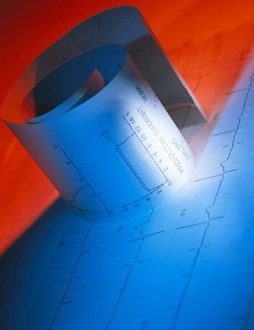
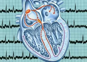
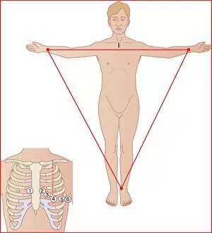
Electrocardiography, commonly known as ECG, is a vital tool in the field of cardiology. It allows medical professionals to monitor and analyze the electrical activity of the human heart. In a normal heart rate, ECGs display a rhythmic pattern resembling artwork on paper. The intricate lines and peaks represent each heartbeat's electrical signals, providing valuable insights into cardiac health. However, not all hearts beat with perfect regularity. Irregularities in heartbeat can be detected through an ECG trace, indicating potential underlying conditions that require attention. The significance lies in its ability to capture the essence of our most vital organ - the human heart. Through computer-generated artwork or real-time monitoring during medical emergencies like an ambulance ride, ECGs provide visual representations that aid cardiologists in diagnosing and treating patients effectively. Sometimes, abnormal patterns such as extrasystole heartbeats appear on an ECG trace. These irregularities are meticulously depicted through artistic interpretations, allowing healthcare professionals to identify potential issues within the cardiovascular system. Cardiology relies heavily on conceptual images derived from electrocardiography for diagnostic purposes. Captivating visuals like C017 / 7718 showcase how this technology merges science and artistry seamlessly. In emergency situations where time is critical, an ambulance becomes a mobile unit equipped with life-saving tools including portable ECG machines. A cardiac patient being monitored en route (C016 / 7443) demonstrates how electrocardiography plays a crucial role in ensuring prompt intervention when every second counts. Whether it's tracing individual beats (C017 / 7744) or capturing overall cardiac function (C017 / 7743), these detailed recordings offer invaluable information about one's cardiovascular health status. Ultimately, electrocardiography stands at the forefront of modern cardiology practices by providing accurate data for diagnosis and treatment plans. Its impact extends beyond medical settings as it continues to inspire awe and fascination through captivating conceptual images (C017 / 7719).