
Endoneurium Collection
The endoneurium is a crucial component of the peripheral nervous system, responsible for supporting and protecting nerve fibers


All Professionally Made to Order for Quick Shipping
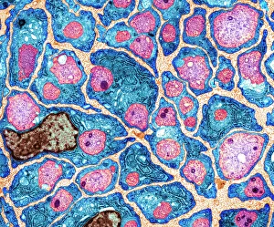
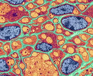
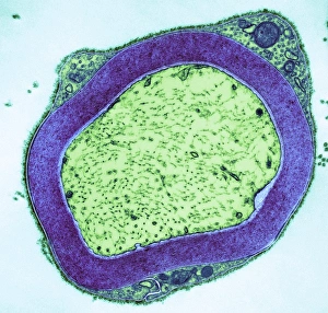
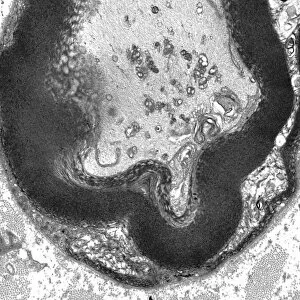
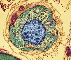
The endoneurium is a crucial component of the peripheral nervous system, responsible for supporting and protecting nerve fibers. Through myelination, these nerve fibers are insulated and able to transmit electrical signals efficiently. In this captivating collection of images captured through transmission electron microscopy (TEM) and light micrograph techniques, we get an up-close look at the intricate details of endoneurium's role in nerve function. In the TEM images, we witness the myelination process in action as thin layers of protective sheaths wrap around individual nerve fibers. These myelinated nerves appear like delicate strands with periodic nodes that enhance signal conduction. The node regions can be observed more closely in separate TEM captures, showcasing their unique structure and importance in maintaining rapid impulse transmission along the fiber. Zooming out from the microscopic world, a light micrograph provides us with a broader view of a peripheral nerve embedded within its surrounding tissue. This image reminds us that while our focus may be on individual components like endoneurium or myelinated nerves, they exist within a complex network that enables communication throughout our body. These stunning visuals serve as reminders of how intricately designed our nervous system is and highlight the significance of structures like endoneurium in ensuring smooth neural communication. As we continue to unravel the mysteries hidden within our own bodies, these glimpses into cellular wonders fuel our curiosity about what lies beneath our skin's surface.