
Endoparasite Collection
"Unveiling the Hidden World of Endoparasites: A Fascinating Journey into Microscopic Intruders" Dog tapeworm head


All Professionally Made to Order for Quick Shipping
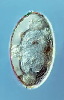
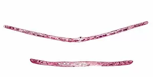
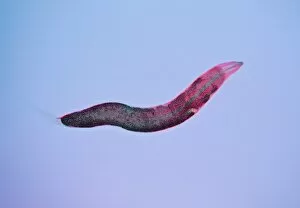
"Unveiling the Hidden World of Endoparasites: A Fascinating Journey into Microscopic Intruders" Dog tapeworm head, SEM: Delving into the intricate anatomy of a dog tapeworm reveals its specialized head structure, designed for attachment and survival within its host. Liver flukes, macro photograph: Zooming in on liver flukes through a macro lens exposes their mesmerizing patterns and shapes, showcasing their ability to adapt and thrive within the liver. Liver fluke egg, macro photograph: Witnessing the minuscule size of a liver fluke egg under close examination highlights how these parasites can reproduce rapidly and infiltrate new hosts. Dog tapeworm, SEM (Picture No. 12479415): This scanning electron microscope image captures the detailed surface texture of a dog tapeworm's body segments, providing insights into its unique adaptations for absorption of nutrients from its host. SEM (Picture No. 12479414): Another stunning SEM image showcases the segmented body structure of a dog tapeworm in all its intricacy – an evolutionary marvel that allows it to grow and reproduce efficiently inside dogs' intestines. SEM (Picture No. 12479416): Capturing yet another perspective with an SEM reveals further details about this endoparasite's morphology – shedding light on how it navigates through various stages of development within its canine host. Parasitized garden tiger caterpillar: Witnessing nature's delicate balance disrupted by endoparasites is both captivating and thought-provoking; here we see a garden tiger caterpillar succumbing to parasitic invasion – reminding us that even seemingly invincible creatures are not immune to microscopic threats. Threadworms in the gut, SEM (Picture No. 12479417).