Fraud Blocker Skip to main content

Endothelial Collection

"Exploring the Intricate World Cells

Background imageEndothelial Collection: Capillary, TEM

Capillary, TEM
Capillary. Transmission electron micrograph (TEM) of a section through a capillary, showing two red blood cells (erythrocytes, black) in its interior

Background imageEndothelial Collection: High endothelial venule, TEM C014 / 1446

High endothelial venule, TEM C014 / 1446
High endothelial venule. Transmission electron micrograph (TEM) of a section through a high endothelial venule (HEV). Venules are tiny veins that transport deoxygenated blood from the capillary beds

Background imageEndothelial Collection: High endothelial venule, TEM C014 / 1445

High endothelial venule, TEM C014 / 1445
High endothelial venule. Transmission electron micrograph (TEM) of a section through a high endothelial venule (HEV). Venules are tiny veins that transport deoxygenated blood from the capillary beds

Background imageEndothelial Collection: Intestinal arteriole, TEM

Intestinal arteriole, TEM
Intestinal arteriole. Transmission electron micrograph (TEM) of a section through an arteriole in the wall of the small intestine. Magnification: x5000 when printed 10 centimetres wide

Background imageEndothelial Collection: Arteriole, TEM

Arteriole, TEM
Arteriole. Transmission electron micrograph (TEM) of a section through a small calibre arterial vessel called an arteriole

Background imageEndothelial Collection: Adrenal gland, fluorescence micrograph C016 / 8472

Adrenal gland, fluorescence micrograph C016 / 8472
Adrenal gland. Fluorescence deconvolution micrograph of a section through an adrenal gland, showing the zona glomerulosa and zona fasciculata

Background imageEndothelial Collection: White blood cell response

White blood cell response
White blood cell movement. Computer-enhanced confocal light micrograph of white blood cells (red) moving through the intact walls of a blood vessel, a process known as diapedesis

Background imageEndothelial Collection: Ovarian blood vessel, SEM

Ovarian blood vessel, SEM
Ovarian blood vessel. Coloured scanning electron micrograph (SEM) of a section through a blood vessel in an ovary. Red blood cells are seen in the lumen, which is lined with endothelial cells (brown)

Background imageEndothelial Collection: High endothelial venule, TEM

High endothelial venule, TEM
High endothelial venule. Coloured transmission electron micrograph (TEM) of the tall cuboidal endothelial cells (upper layer) lining a high endothelial venule (HEV)

Background imageEndothelial Collection: Heart blood vessels, SEM

Heart blood vessels, SEM
Heart blood vessels. Coloured scanning electron micrograph (SEM) of a freeze-fractured section through cardiac muscle, exposing the blood vessels of the heart

Background imageEndothelial Collection: Artery anatomy, artwork

Artery anatomy, artwork
Artery anatomy, computer artwork. At the centre of the artery is the lumen, with three red blood cells (erythrocytes, bottom right)

Background imageEndothelial Collection: Arteriole and red blood cells, SEM

Arteriole and red blood cells, SEM
Arteriole. Coloured scanning electron micrograph (SEM) of a cross-section through a small, thin- walled artery known as an arteriole

Background imageEndothelial Collection: Brain artery, SEM

Brain artery, SEM
Brain artery. Coloured scanning electron micrograph (SEM) of a freeze-fractured artery from the brain. The fracture plane has passed through the artery

Background imageEndothelial Collection: Stressed culture cell, SEM

Stressed culture cell, SEM

Background imageEndothelial Collection: Heart ventricle, SEM

Heart ventricle, SEM
Heart ventricle. Coloured scanning electron micrograph (SEM) of a fracture through a heart to show the interior of the ventricle. Erythrocytes (red blood cells) are seen within the ventricle

Background imageEndothelial Collection: Blood vessel repair, artwork

Blood vessel repair, artwork
Blood vessel repair. Computer artwork showing the various cellular mechanisms and interactions involved in the repair of blood vessels after damage.See image: C007/8662 for the labelled diagram

Background imageEndothelial Collection: Asthma pathology, artwork

Asthma pathology, artwork
Asthma pathology. Computer artwork showing cross-sections through a normal airway (right) and an asthmatic, constricted airway (left)

Background imageEndothelial Collection: Lung cells, fluorescent micrograph

Lung cells, fluorescent micrograph
Lung cells. Immunofluorescence light micrograph of pulmonary endothelial cells. Endothelial cells are specialized epithelial cells that line the inner surface of blood vessels


All Professionally Made to Order for Quick Shipping

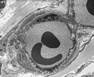
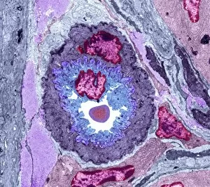
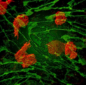
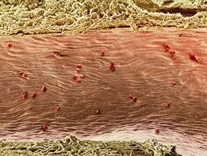
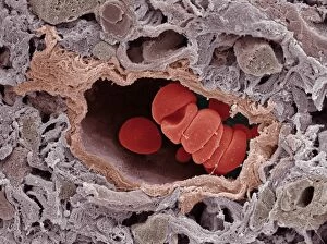
"Exploring the Intricate World Cells: A Glimpse through TEM" Captivating images captured under a transmission electron microscope (TEM) reveal the fascinating world cells. In one snapshot, we witness the intricate dance between capillaries and red blood cells, as they navigate through the complex network of vessels. The TEM allows us to delve deeper into this microscopic realm, providing unprecedented details. The first image showcases a close-up view of a capillary and red blood cell interaction. Through TEM, we can observe their delicate connection and understand how these tiny vessels facilitate oxygen exchange within our bodies. This remarkable insight opens new avenues for studying cardiovascular health and disease prevention. Moving on to another captivating capture, we encounter high endothelial venules (HEVs), depicted in stunning detail by TEM C014 / 1446. These specialized postcapillary venules play a crucial role in immune responses by enabling lymphocyte migration from the bloodstream into lymphoid organs. The intricacies revealed by this image shed light on the body's defense mechanisms at an astonishing level. Intriguingly, our exploration continues with yet another glimpse into HEVs using TEM C014 / 1445. This particular image offers further insights into their structure and function, unraveling mysteries surrounding immune cell trafficking during inflammation or infection processes. Shifting focus towards intestinal arterioles, we are presented with three striking images captured via TEM. These snapshots provide an up-close look at these small arteries that supply vital nutrients to our intestines while maintaining proper blood flow regulation. Such detailed visualization aids researchers in understanding digestive disorders and designing targeted therapies. As we conclude our journey through these mesmerizing TEM images cells, it becomes evident that unlocking their secrets holds immense potential for advancing medical knowledge and improving human health outcomes. From unravelling cellular interactions within capillaries to deciphering immune responses mediated by HEVs, these microscopic wonders continue to captivate and inspire scientists worldwide.