Fraud Blocker Skip to main content

Epidermis Collection (page 8)

The epidermis, our body's protective outer layer, is a fascinating subject that encompasses various aspects of skin disorders and artwork

Background imageEpidermis Collection: Powdery scab infection, light micrograph

Powdery scab infection, light micrograph
Powdery scab infection. Light micrograph of a section through a potato infected with powdery scab (Spongospora subterranea), showing pustules and spores. S

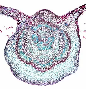
Background imageEpidermis Collection: Sycamore leaf vein, light micrograph

Sycamore leaf vein, light micrograph
Sycamore leaf vein. Light micrograph of a section through the midrib (vein) of a leaf from a sycamore (Acer pseudoplatanus) tree

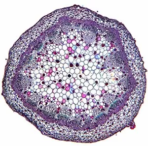
Background imageEpidermis Collection: Geranium stem, light micrograph

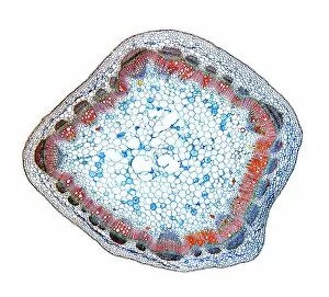
Geranium stem, light micrograph
Geranium stem. Light micrograph of a section through a young stem of a geranium (Pelargonium sp.) plant. The outer stem is covered with a thin epidermis (red) which has stomata

Background imageEpidermis Collection: Sweet pea stem, light micrograph

Sweet pea stem, light micrograph
Sweet pea stem. Light micrograph of a section through the hollow stem of a sweet pea (Lathyrus odoratus) plant, showing a ring of vascular bundles

Background imageEpidermis Collection: Rust fungus in a leaf, light micrograph

Rust fungus in a leaf, light micrograph
Rust fungus in a leaf. Light micrograph of a section through a wheat leaf infected with the parasitic Puccinia graminis rust

Background imageEpidermis Collection: Sage stem, light micrograph

Sage stem, light micrograph
Sage stem. Light micrograph of a section through a primary stem of a scarlet sage (Salivia splendens) plant. The outer stem is covered with a thin epidermis (green) that contains stomata

Background imageEpidermis Collection: Lime tree stem wound, light micrograph

Lime tree stem wound, light micrograph
Lime tree stem wound. Light micrograph of a section through the wounded stem of a lime tree (Tilia europaea). The outer epidermis has been shed and replaced by a layer of cork (dark red)

Background imageEpidermis Collection: Beech tree leaves, light micrograph

Beech tree leaves, light micrograph
Beech tree leaves. Light micrograph of a section through two leaves from different parts of a common beech tree (Fagus sylvatica)

Background imageEpidermis Collection: Hollyhock rust fungus in a leaf

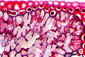
Hollyhock rust fungus in a leaf. Light micrograph of a section through a hollyhock (Alcea sp.) leaf infected with the parasitic Puccinia malvacearum rust, showing the teliospores (spores)

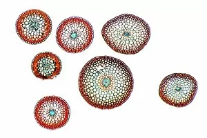
Background imageEpidermis Collection: Horse-tail stem, light micrograph

Horse-tail stem, light micrograph
Rice grass stem. Light micrograph of a section through a rice grass stem (Oryza sativa). Vascular bundles (small oval structures) can be seen containing xylem (larger openings)

Background imageEpidermis Collection: Oleander leaf, light micrograph

Oleander leaf, light micrograph
Oleander leaf. Light micrograph of a section through the leaf of an oleander (Nerium oleander) tree, showing its sunken stomata (gaps, centre left and right)

Background imageEpidermis Collection: Plant root development, artwork

Plant root development, artwork
Plant root development. Artwork showing the first three stages in the development of a plant root. The first of these three stages (upper left)

Background imageEpidermis Collection: Plant root development, diagram

Plant root development, diagram
Plant root development. Diagram showing the the fourth (left) and fifth (right) stages in a series showing the development of a plant root

Background imageEpidermis Collection: Southern elephant seal moulting

Southern elephant seal moulting
Southern elephant seal (Mirounga leonina) juvenile moulting. Southern elephant seals live in the open waters of the Southern Ocean around Antarctica

Background imageEpidermis Collection: Annelid worm anatomy

Annelid worm anatomy. Computer artwork showing the internal anatomy of 3 segments (metameres) from an annelid (segmented) worms body

Background imageEpidermis Collection: Moss stems, light micrograph

Moss stems, light micrograph. Transverse section through stems of a cord moss (Polytrichum commune). The outside of the stems, the epidermis, is made up of thick-walled cells

Background imageEpidermis Collection: Fern stem, light micrograph

Fern stem, light micrograph. Transverse section through a rachis (stem) of the bracken fern (Pteridium aquilinum). Under the outer epidermis (black) is a thin cortex (deep red)

Background imageEpidermis Collection: Horsetail stem, light micrograph

Horsetail stem, light micrograph. Transverse section through the stem of the common horsetail (Equisetum arvensis). The stem consists of an outer epidermis and an outer cortex of sclerenchyma cells

Background imageEpidermis Collection: Hair follicle, SEM

Hair follicle, SEM
Hair follicle. Coloured scanning electron micrograph (SEM) of an empty hair follicle (centre) among squamous cells on the surface of the skin

Background imageEpidermis Collection: Skin cells, SEM

Skin cells, SEM
Skin cells. Coloured scanning electron micrograph (SEM) of squamous cells from the surface of the skin. These are flat, keratinised

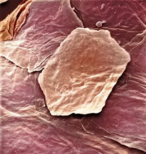
Background imageEpidermis Collection: Skin cell, SEM

Skin cell, SEM
Skin cell. Coloured scanning electron micrograph (SEM) of a squamous cell on the surface of the skin. This is a flat, keratinised, dead cell

Background imageEpidermis Collection: Human skin surface, SEM

Human skin surface, SEM
Human skin surface, coloured scanning electron micrograph (SEM). Ridges and loose squamous cells on the surface of the skin

Background imageEpidermis Collection: Leaf section, SEM

Leaf section, SEM
Leaf section. Coloured scanning electron micrograph (SEM) of a section through a fractured leaf. At top is a single layer of cells that forms the epidermis of the leaf

Background imageEpidermis Collection: Flax plant stem, light micrograph

Flax plant stem, light micrograph
Flax plant stem. Light micrograph of a transverse section through a stem of the flax plant (Linum usitatissimum). The layers from outer to inner (some very thin) are the epidermis (bottom)

Background imageEpidermis Collection: Peanut plant stem, light micrograph

Peanut plant stem, light micrograph
Peanut plant stem. Light micrograph of a transverse section through a stem of the peanut plant (Arachis hypogaea). Below the stems outer layer (epidermis) is a cortex of parenchyma cells (blue)

Background imageEpidermis Collection: Stinging nettle stem, light micrograph

Stinging nettle stem, light micrograph
Stinging nettle stem. Polarised light micrograph of a transverse section through a stem of the stinging nettle plant (Urtica dioica)

Background imageEpidermis Collection: Tea leaf, light micrograph

Tea leaf, light micrograph
Tea leaf. Light micrograph of a cross-section through a tea (Camellia sinensis) leaf. The upper and lower epidermis on the surfaces of the leaf are blue

Background imageEpidermis Collection: Pine needle, light micrograph

Pine needle, light micrograph
Pine needle. Light micrograph of a transverse section through a leaf (needle) of a pine tree (Pinus sp.). The leaves are needle-like so they present a large surface area for photosynthesis but

Background imageEpidermis Collection: Broad bean root, light micrograph

Broad bean root, light micrograph
Broad bean root. Light micrograph of a transverse section through the young root of a broad bean (Vicia faba) plant. The outer epidermis is covered in root hairs

Background imageEpidermis Collection: Kidney bean root, light micrograph

Kidney bean root, light micrograph
Kidney bean root. Light micrograph of a section through the root of a kidney bean (Phaseolus vulgaris) plant. At centre is the pith (blue)

Background imageEpidermis Collection: Common broom stem, light micrograph

Common broom stem, light micrograph
Common broom stem. Light micrograph of a transverse section through the stem of a common broom (Salicornia europaea) plant

Background imageEpidermis Collection: Young pine tree stem, light micrograph

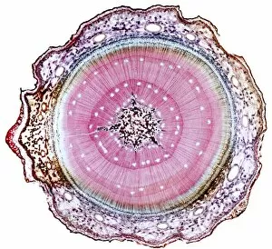
Young pine tree stem, light micrograph
Young pine tree stem. Light micrograph of a transverse section through a two and a half-year-old stem of a pine tree (Pinus sp.)

Background imageEpidermis Collection: Dyers greenweed stem, light micrograph

Dyers greenweed stem, light micrograph
Dyers greenweed stem. Polarised light micrograph of a transverse section through the stem of a dyers greenweed (Genista tinctoria) plant

Background imageEpidermis Collection: Marsh samphire stem, light micrograph

Marsh samphire stem, light micrograph
Marsh samphire stem. Light micrograph of a transverse section through the stem of a marsh samphire, (Salicornia europaea) plant. This is a succulent, xerophytic, halophyte plant

Background imageEpidermis Collection: Ginkgo leaf stalk, light micrograph

Ginkgo leaf stalk, light micrograph
Ginkgo leaf stalk. Light micrograph of a transverse section through the leaf stalk (petiole) of a ginkgo tree (Ginkgo biloba)

Background imageEpidermis Collection: Cedar tree stem, light micrograph

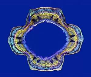
Cedar tree stem, light micrograph
Cedar tree stem. Light micrograph of a transverse section through a stem of a cedar tree (Thujopsis dolobrata). The four ridges on the outer surface are microphyllous leaves

Background imageEpidermis Collection: Sunflower stem, light micrograph

Sunflower stem, light micrograph
Sunflower stem. Light micrograph of a transverse section through the stem of a sunflower (Helianthus annuus) plant, showing a vascular bundle

Background imageEpidermis Collection: Elder tree stem, light micrograph

Elder tree stem, light micrograph
Elder tree stem. Light micrograph of a transverse section through the very young woody stem of an elder (Sambuca nigra) tree

Background imageEpidermis Collection: Japanese sago palm leaf, light micrograph

Japanese sago palm leaf, light micrograph
Japanese sago palm leaf. Light micrograph of a transverse section through the leaf (pinna) of a Japanese sago palm, cycad, (Cycas revoluta)

Background imageEpidermis Collection: Sunflower leaf, light micrograph

Sunflower leaf, light micrograph
Sunflower leaf. Light micrograph of a transverse section through the midrib of a sunflower (Helianthus annuus) leaf. The upper and lower epidermis on the surfaces of the leaf are blue

Background imageEpidermis Collection: Eucalyptus stem, light micrograph

Eucalyptus stem, light micrograph
Eucalyptus stem. Light micrograph of a transverse section through a one-year-old stem of a Eucalyptus (Eucalyptus globulus) plant

Background imageEpidermis Collection: Yew tree leaf, light micrograph

Yew tree leaf, light micrograph
Yew tree leaf. Light micrograph of a transverse section through the leaf (pinna) of a yew tree, (Taxus baccata). The structure has xerophytic (drought plants) characteristics


All Professionally Made to Order for Quick Shipping

The epidermis, our body's protective outer layer, is a fascinating subject that encompasses various aspects of skin disorders and artwork. From examining the lime tree stem under a light micrograph to exploring a section through human skin using scanning electron microscopy (SEM), there are endless possibilities for understanding this intricate organ. In Picture No. 11675585, we can observe an illustration depicting the cross-section of human skin with heat trapped by erect hairs. This visual representation highlights the complexity of our epidermis and its role in regulating body temperature. Delving deeper into the layers of our skin, SEM images reveal the intricate patterns and structures present on its surface. Whether it's studying the texture of tomato leaf or periwinkle petal surfaces or even analyzing zebra fish skin at a microscopic level, these images provide valuable insights into how diverse organisms have adapted their epidermal layers for survival. Examining individual cells within the epidermis becomes possible through transmission electron microscopy (TEM). By zooming in on these tiny building blocks, we gain insight into their structure and function, helping us understand how they contribute to overall skin health. Even beyond humans, other organisms like olive leaves also possess unique features worth exploring. Through SEM imaging of olive leaf trichomes - small hair-like structures covering their surface - we can appreciate nature's intricacies while drawing parallels between different species' adaptations. From understanding common skin disorders to appreciating art inspired by this remarkable organ's beauty and functionality, delving into the world opens up a realm filled with scientific wonder and artistic inspiration alike.