Fraud Blocker Skip to main content

Epithelial Collection

Epithelial cells, the unsung heroes of our body's lining

Background imageEpithelial Collection: Intestinal lining

Intestinal lining

Background imageEpithelial Collection: Trachea lining, SEM

Trachea lining, SEM
Trachea lining, coloured scanning electron micrograph (SEM). The tracheal lining contains mucus-secreting goblet cells and epithelial cells bearing hair-like cilia (blue)

Background imageEpithelial Collection: Intestinal microvilli, TEM

Intestinal microvilli, TEM
Intestinal microvilli, coloured transmission electron micrograph (TEM). The microvilli are finger-like projections (seen here in circular cross-section)

Background imageEpithelial Collection: Destruction complete d une moitie du visage par un cancer epithelial (colour litho)

Destruction complete d une moitie du visage par un cancer epithelial (colour litho)
5210424 Destruction complete d une moitie du visage par un cancer epithelial (colour litho) by French School, (19th century); Private Collection; (add.info)

Background imageEpithelial Collection: Cross-section diagram of a cancerous tumour including calcium deposits, blood vessels

Cross-section diagram of a cancerous tumour including calcium deposits, blood vessels, tumour outgrowth, epithelial layer, ulcerated area, bleeding, nerve fibres, dead tissue and a lymph vessel

Background imageEpithelial Collection: Nasal lining, SEM

Nasal lining, SEM
Nasal lining. Coloured scanning electron micrograph (SEM) of the olfactory epithelium that lines the nasal cavity, showing olfactory cells (red) surrounded by numerous cilia (hair-like projections)

Background imageEpithelial Collection: Intestinal villi, SEM

Intestinal villi, SEM
Intestinal villi. Coloured scanning electron micrograph (SEM) of a section through a part of the small intestine, showing the villi that cover its inner surface

Background imageEpithelial Collection: Throat cancer, X-rays

Throat cancer, X-rays
Throat cancer. Coloured X-rays showing the transit of a contrast medium (dense white) through the throat of a 54-year-old patient with an adenocarcinoma (cancer) of the lower oesophagus (gullet)

Background imageEpithelial Collection: Trachea lining, SEM C015 / 9936

Trachea lining, SEM C015 / 9936
Trachea lining. Coloured scanning electron micrograph (SEM) of a section through the lining of the trachea (wind pipe). The trachea links the larynx (voice box) to the lungs

Background imageEpithelial Collection: Colon cancer, endoscopic view C016 / 8308

Colon cancer, endoscopic view C016 / 8308
Colon cancer. Endoscopic view of a malignant (cancerous) adenocarcinoma (upper right) in a patients ascending colon (large intestine)

Background imageEpithelial Collection: Trachea lining, SEM C013 / 7126

Trachea lining, SEM C013 / 7126
Trachea lining. Coloured scanning electron micrograph (SEM) of the lining of a trachea (wind pipe), showing the cilia (hair-like) and goblet cells (orange). The trachea links the larynx to the lungs

Background imageEpithelial Collection: Trachea lining, SEM C013 / 7122

Trachea lining, SEM C013 / 7122
Trachea lining. Coloured scanning electron micrograph (SEM) of a section through the wall of a trachea (wind pipe). The trachea links the larynx to the lungs

Background imageEpithelial Collection: Bladder epithelium, light micrograph

Bladder epithelium, light micrograph
Bladder epithelium. Light micrograph of a vertical section through the wall of the urinary bladder. The inner surface is at top

Background imageEpithelial Collection: Trachea, SEM

Trachea, SEM
Trachea. Coloured scanning electron micrograph (SEM) of the lining of the trachea (windpipe). The trachea links the larynx (voice box) to the lungs

Background imageEpithelial Collection: Conceptual image of the Human Papilloma Virus

Conceptual image of the Human Papilloma Virus (HPV). HPV is a virus from the papillomavirus family that affects human skin and the moist membranes that line the body

Background imageEpithelial Collection: Trachea lining, SEM C016 / 9756

Trachea lining, SEM C016 / 9756
Trachea lining. Coloured scanning electron micrograph (SEM) of the lining of the trachea (wind pipe). The trachea links the larynx to the lungs

Background imageEpithelial Collection: Sunburnt skin

Sunburnt skin. Coloured electron micrograph (SEM) of epidermal skin cells after sunburn. These stratified epithelial cells have been destroyed by ultraviolet (UV)

Background imageEpithelial Collection: Human skin, SEM

Human skin, SEM
Human skin. Coloured scanning electron micrograph (SEM) of the surface layer of the human skin, the epidermis. The outer layer of the epidermis (the stratum corneum)

Background imageEpithelial Collection: Bladder lining, TEM C014 / 1473

Bladder lining, TEM C014 / 1473
Bladder lining. Transmission electron micrograph (TEM) of a section through the epithelial cells that line of a human bladder, showing large flat vesicles (large, blue)

Background imageEpithelial Collection: Stem cell-derived retinal cells

Stem cell-derived retinal cells. Fluorescence light micrograph of retinal pigment epithelium (RPE) cells that have been derived from human embryonic stem cells (HESC)

Background imageEpithelial Collection: Breast cancer, TEM F006 / 8633

Breast cancer, TEM F006 / 8633
Breast cancer. Coloured transmission electron micrograph (TEM) of a section through a ductal carcinoma (cancer) of the breast showing malignant epithelial cells

Background imageEpithelial Collection: Breast cancer, TEM F006 / 8632

Breast cancer, TEM F006 / 8632
Breast cancer. Coloured transmission electron micrograph (TEM) of a section through a ductal carcinoma (cancer) of the breast showing malignant epithelial cells

Background imageEpithelial Collection: Colitis, light micrograph

Colitis, light micrograph
Colitis. Light micrograph of a section through colon (large intestine) tissue affected by bacterial infection, causing injury to the mucosal lining, which has resulted in inflammation (colitis)

Background imageEpithelial Collection: Hairy scalp skin, light micrograph

Hairy scalp skin, light micrograph
Hairy scalp skin. Light micrograph of a section through healthy skin from a human scalp, showing the numerous hair follicles. The scalp is an example of hairy skin

Background imageEpithelial Collection: Ischaemic bowel, light micrograph

Ischaemic bowel, light micrograph
Ischaemic bowel. Light micrograph of a section through a portion of intestine that has been damaged by an interruption of the blood supply, or ischaemia

Background imageEpithelial Collection: Staphylococcus aureus bacteria, TEM C016 / 9458

Staphylococcus aureus bacteria, TEM C016 / 9458
Staphylococcus aureus bacteria. Coloured transmission electron micrograph (TEM) of a Staphylococcus aureus bacterium (round) in skin tissue. S

Background imageEpithelial Collection: Helicobacter pylori bacteria, SEM C016 / 9135

Helicobacter pylori bacteria, SEM C016 / 9135
Helicobacter pylori bacteria. Coloured scanning electron micrograph (SEM) of Helicobacter pylori bacteria (yellow) in the stomach. H

Background imageEpithelial Collection: Helicobacter pylori bacteria, SEM C016 / 9134

Helicobacter pylori bacteria, SEM C016 / 9134
Helicobacter pylori bacteria. Coloured scanning electron micrograph (SEM) of Helicobacter pylori bacteria (orange) in the stomach. H

Background imageEpithelial Collection: Helicobacter pylori bacteria, SEM C016 / 9136

Helicobacter pylori bacteria, SEM C016 / 9136
Helicobacter pylori bacteria. Coloured scanning electron micrograph (SEM) of Helicobacter pylori bacteria (green) in the stomach. H

Background imageEpithelial Collection: Staphylococcus aureus bacteria, TEM C016 / 9457

Staphylococcus aureus bacteria, TEM C016 / 9457
Staphylococcus aureus bacteria. Coloured transmission electron micrograph (TEM) of a Staphylococcus aureus bacterium (blue) in skin tissue. S

Background imageEpithelial Collection: Staphylococcus aureus bacteria, TEM C016 / 9455

Staphylococcus aureus bacteria, TEM C016 / 9455
Staphylococcus aureus bacteria. Coloured transmission electron micrograph (TEM) of Staphylococcus aureus bacteria four hours after infection of pulmonary epithelial cells. S

Background imageEpithelial Collection: Helicobacter pylori bacteria, SEM C016 / 9053

Helicobacter pylori bacteria, SEM C016 / 9053
Helicobacter pylori bacteria. Coloured scanning electron micrograph (SEM) of a Helicobacter pylori bacterium (blue) in the stomach. H

Background imageEpithelial Collection: Staphylococcus aureus bacteria, TEM C016 / 9456

Staphylococcus aureus bacteria, TEM C016 / 9456
Staphylococcus aureus bacteria. Coloured transmission electron micrograph (TEM) of Staphylococcus aureus bacteria four hours after infection of pulmonary epithelial cells. S

Background imageEpithelial Collection: Finger skin, SEM

Finger skin, SEM
Finger skin. Coloured scanning electron micrograph (SEM) of a section through skin from a human finger, showing the characteristic dermal ridges (lower left, and right) that make up the fingerprint

Background imageEpithelial Collection: Ductal breast cancer, TEM

Ductal breast cancer, TEM
Ductal breast cancer. Transmission electron micrograph (TEM) of a section through breast tissue, showing malignant (cancerous) epithelial cells arising from the milk ducts of the breast

Background imageEpithelial Collection: Human Skin Cells (SEM)

Human Skin Cells (SEM)
Human Skin Cells. Scanning electron micrograph (SEM) of the surface layer of the human skin, the epidermis. The outer layer of the epidermis (stratum corneum)

Background imageEpithelial Collection: Smell receptor, TEM

Smell receptor, TEM
Smell receptor. Transmission electron micrograph (TEM) of a section through the olfactory epithelium that lines the nasal cavity, showing an olfactory cell (smell receptor)

Background imageEpithelial Collection: Goblet cell, TEM

Goblet cell, TEM
Goblet cell. Transmission electron micrograph (TEM) of a section through a goblet cell in the lining (epithelium, top) of the small intestine

Background imageEpithelial Collection: Zebra fish skin, SEM

Zebra fish skin, SEM
Zebra fish skin. Coloured scanning electron micrograph (SEM) of the folded surface of skin cells from a zebrafish (Danio rerio). The microscopic folds are called microplicae

Background imageEpithelial Collection: Intestinal brush border, TEM

Intestinal brush border, TEM
Intestinal brush border. Transmission electron micrograph (TEM) of intestinal absorptive cells sectioned horizontally at their apex to show the surface microvilli (round)

Background imageEpithelial Collection: Trachea lining, SEM C015 / 9939

Trachea lining, SEM C015 / 9939
Trachea lining. Coloured scanning electron micrograph (SEM) of a section through the lining of the trachea (wind pipe). The trachea links the larynx (voice box) to the lungs

Background imageEpithelial Collection: Gastritis, light micrograph C016 / 0508

Gastritis, light micrograph C016 / 0508
Gastritis. Light micrograph of a section through an inflamed human stomach (gastritis), showing an abnormal accumulation of white blood cells (leucocytes)

Background imageEpithelial Collection: Trachea lining, SEM C015 / 9610

Trachea lining, SEM C015 / 9610
Trachea lining. Coloured scanning electron micrograph (SEM) of the lining of the trachea (wind pipe). The trachea links the larynx to the lungs

Background imageEpithelial Collection: Retina of the eye, light micrograph C016 / 0528

Retina of the eye, light micrograph C016 / 0528
Retina of the eye. Light micrograph of a section through the retina from a human eye. From top down: nerve fibres of the optic nerve and a blood vessel; several layers of neurons (nerve cells)

Background imageEpithelial Collection: Gastritis, light micrograph C016 / 0507

Gastritis, light micrograph C016 / 0507
Gastritis. Light micrograph of a section through an inflamed human stomach (gastritis), showing an abnormal accumulation of white blood cells (leucocytes)

Background imageEpithelial Collection: Lens of the eye, light micrograph C016 / 0526

Lens of the eye, light micrograph C016 / 0526
Lens of the eye. Light micrograph of a section through the lens from a human eye. The lens consists of thousands of precisely aligned stacks of fibres seen here end-on

Background imageEpithelial Collection: Stratified squamous epithelium C016 / 0534

Stratified squamous epithelium C016 / 0534
Stratified squamous epithelium. Light micrograph of a section through stratified squamous epithelium, showing the multiple layers, or strata, that function to resist abrasion of the surface

Background imageEpithelial Collection: Gallbladder surface, SEM C015 / 9613

Gallbladder surface, SEM C015 / 9613
Gallbladder surface, coloured scanning electron micrograph (SEM). The columnar epithelium of the surface is covered in microvilli (tiny hair-like projections)


All Professionally Made to Order for Quick Shipping

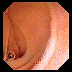
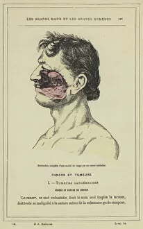
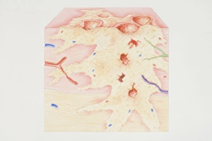
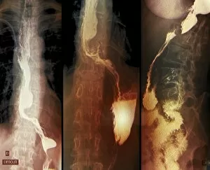
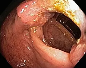
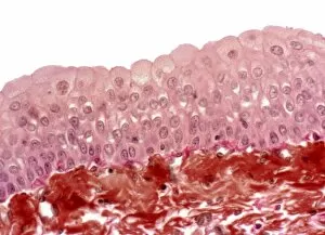
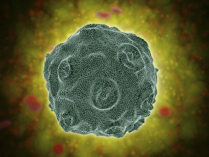
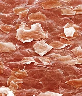
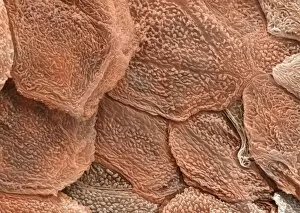
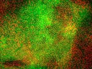
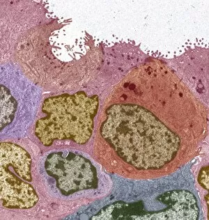
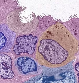
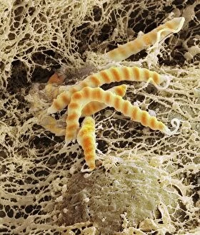
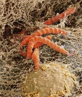
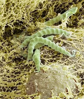
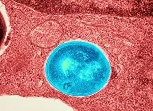
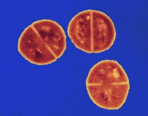
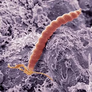
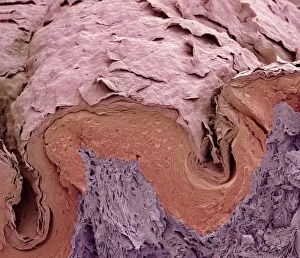
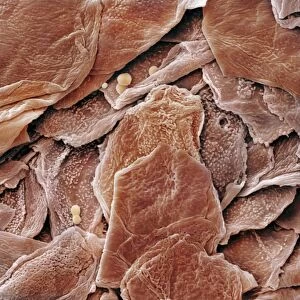
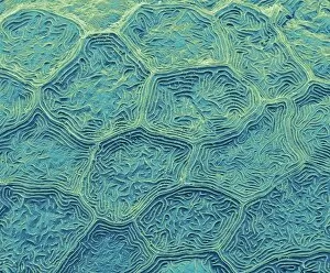
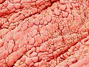
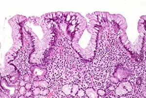
Epithelial cells, the unsung heroes of our body's lining. From the intestinal tract to the trachea, these microscopic warriors protect and support us in ways we often overlook. Take a closer look at the intestinal lining under a scanning electron microscope (SEM). The intricate network of microvilli reveals itself, enhancing nutrient absorption and ensuring our digestive system functions seamlessly. Moving on to the trachea lining, SEM unveils its unique structure. These epithelial cells form a protective barrier against harmful particles while allowing for efficient gas exchange within our respiratory system. Zooming into zebra fish skin through SEM, we discover another fascinating aspect cells. Their arrangement provides defense against external threats while maintaining essential functions like osmoregulation. Intriguingly captured by SEM are bacteria residing in our nasal passages. Though seemingly insignificant, these tiny organisms play crucial roles in maintaining healthy immune responses and preventing infections. However, not all stories about epithelial cells are positive. A colour lithograph depicts the devastating impact of cancerous growth on half of someone's face. This serves as a reminder that even resilient epithelial layers can succumb to destructive forces if left unchecked. A cross-section diagram further illustrates how cancer affects various components such as calcium deposits, blood vessels, ulcerated areas, nerve fibers - all intertwined with an afflicted epithelial layer. It emphasizes the importance of early detection and treatment options available for those battling this formidable disease. Returning to less dire scenarios captured by SEM is the nasal lining – yet another example showcasing how delicate but vital these cellular linings truly are. The intestinal villi also make their appearance under SEM – finger-like projections that increase surface area for optimal nutrient absorption within our gut ecosystem. Shifting gears towards throat cancer diagnosis using X-rays highlights how medical imaging techniques aid in identifying abnormalities affecting this critical part of our anatomy. Early detection can significantly improve prognosis and treatment outcomes for patients facing such challenges.