Fraud Blocker Skip to main content

Erythrocyte Collection (page 3)

"Erythrocyte: The Lifeline of Blood" Erythrocytes, commonly known as red blood cells, play a crucial role in our circulatory system

Background imageErythrocyte Collection: Microscopic view of human anitbodies with red blood cells

Microscopic view of human anitbodies with red blood cells. Human antibodies are the Y-shaped proteins used by the immune system to defend against foreign objects like bacteria and viruses
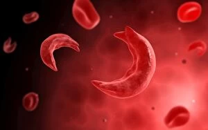
Background imageErythrocyte Collection: Microscopic view of sicke cells causing anemia disease

Microscopic view of sicke cells causing anemia disease

Background imageErythrocyte Collection: Microscopic view of blood cells

Microscopic view of blood cells

Background imageErythrocyte Collection: Conceptual image of a blood vessel

Conceptual image of a blood vessel

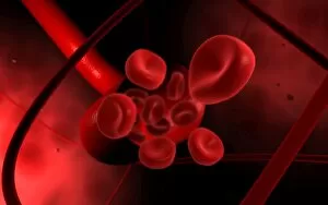
Background imageErythrocyte Collection: Conceptual image of red blood cells

Conceptual image of red blood cells

Background imageErythrocyte Collection: Components of blood vessel interact with intracellular fluid

Components of blood vessel interact with intracellular fluid; white blood cells, red blood cells, oxygen, platelets and hormones

Background imageErythrocyte Collection: Malria spores in the human blood stream

Malria spores in the human blood stream
The malaria spores move from the mosquito to the human blood stream. the spores divide many times and produce large numbers of spores

Background imageErythrocyte Collection: Microscopic view of red blood cells flowing inside lungs

Microscopic view of red blood cells flowing inside lungs

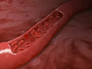
Background imageErythrocyte Collection: Artery cross section with red blood cell flow

Artery cross section with red blood cell flow

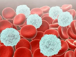
Background imageErythrocyte Collection: Red blood cells with white blood cells

Red blood cells with white blood cells

Background imageErythrocyte Collection: Blood clot, SEM C016 / 9751

Blood clot, SEM C016 / 9751
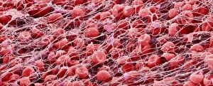
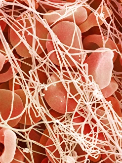
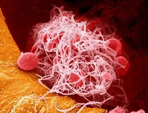
Blood clot, coloured scanning electron micrograph (SEM). Red blood cells (erythrocytes) are trapped within a fibrin protein mesh (beige)

Background imageErythrocyte Collection: Blood clot, SEM C016 / 9746

Blood clot, SEM C016 / 9746
Blood clot, coloured scanning electron micrograph (SEM). Red blood cells (erythrocytes) are trapped within a fibrin protein mesh (beige)

Background imageErythrocyte Collection: Blood clot, SEM C016 / 9749

Blood clot, SEM C016 / 9749
Blood clot, coloured scanning electron micrograph (SEM). Red blood cells (erythrocytes) are trapped within a fibrin protein mesh (beige)

Background imageErythrocyte Collection: Blood clot, SEM C017 / 7141

Blood clot, SEM C017 / 7141
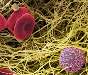
Blood clot, coloured scanning electron micrograph (SEM). Red blood cells (erythrocytes) and a white blood cell (purple) trapped within a fibrin protein mesh (cream)

Background imageErythrocyte Collection: Blood clot, SEM C016 / 9750

Blood clot, SEM C016 / 9750
Blood clot, coloured scanning electron micrograph (SEM). Red blood cells (erythrocytes) are trapped within a fibrin protein mesh (beige)

Background imageErythrocyte Collection: Blood clot, SEM C016 / 9753

Blood clot, SEM C016 / 9753
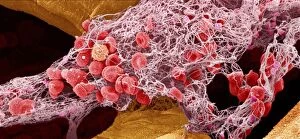
Blood clot. Coloured scanning electron micrograph (SEM) of a blood clot from the inner wall of the left ventricle of a heart

Background imageErythrocyte Collection: Blood clot, SEM C016 / 9752

Blood clot, SEM C016 / 9752
Blood clot, coloured scanning electron micrograph (SEM). Red blood cells (erythrocytes) are trapped within a fibrin protein mesh (beige)

Background imageErythrocyte Collection: Red blood cells, artwork F008 / 0202

Red blood cells, artwork F008 / 0202
Red blood cells, computer artwork

Background imageErythrocyte Collection: Blood clot, SEM P260 / 0123

Blood clot, SEM P260 / 0123
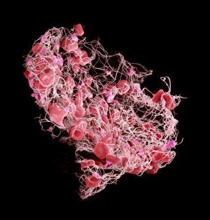
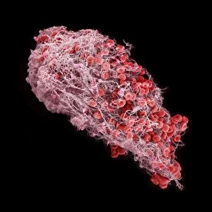
Blood clot. Coloured scanning electron micrograph (SEM) of a blood clot. The red blood cells (erythrocytes) are trapped in filaments of fibrin protein (pink)

Background imageErythrocyte Collection: Colour SEM of red & white blood cells

Colour SEM of red & white blood cells
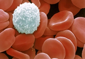
Blood cells. Coloured scanning electron micrograph (SEM) of human blood showing red and white cells. Red blood cells (erythrocytes) have a characteristic biconcave-disc shape and are numerous

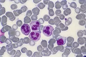
Background imageErythrocyte Collection: Blood smear, light micrograph F005 / 6090

Blood smear, light micrograph F005 / 6090
Blood smear. Light micrograph showing normal red and white blood cells. At upper centre is monocyte, at top right and centre right are neutrophils, at bottom right is a basoophil

Background imageErythrocyte Collection: Blood clot, SEM C016 / 9745

Blood clot, SEM C016 / 9745
Blood clot, coloured scanning electron micrograph (SEM). Red blood cells (erythrocytes) are trapped within a fibrin protein mesh (beige)

Background imageErythrocyte Collection: Blood clot, SEM C016 / 9748

Blood clot, SEM C016 / 9748
Blood clot, coloured scanning electron micrograph (SEM). Red blood cells (erythrocytes) are trapped within a fibrin protein mesh (beige)

Background imageErythrocyte Collection: Immunotherapy, artwork

Immunotherapy, artwork
Immunotherapy. Computer artwork of immunotherapy being used to destroy a tumour (right). Immunotherapy uses the bodys immune system to fight a disease

Background imageErythrocyte Collection: Haematopoietic stem cells, artwork

Haematopoietic stem cells, artwork
Haematopoietic stem cells. Cutaway computer artwork showing white blood cells (leucocytes, white, round), red blood cells (erythrocytes, red) and haematopoietic stem cells (HSCs)

Background imageErythrocyte Collection: Crenated red blood cells, SEM C016 / 9029

Crenated red blood cells, SEM C016 / 9029
Crenated red blood cells. Coloured scanning electron micrograph (SEM) of a section through an arteriole (small blood vessel), showing crenated red blood cells inside

Background imageErythrocyte Collection: Human bone structure, artwork C016 / 7494

Human bone structure, artwork C016 / 7494
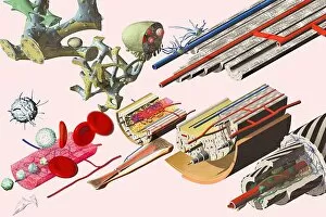
Human bone structure. Computer artwork showing all the different parts that make up a human bone, including the bone marrow (bottom left) where blood cells (round, white and red) are formed

Background imageErythrocyte Collection: Atherosclerosis in artery, artwork C016 / 6571

Atherosclerosis in artery, artwork C016 / 6571
Atherosclerosis in artery. Artwork of a longitudinal section through an artery that has been narrowed by atherosclerosis. Narrowing is referred to as stenosis

Background imageErythrocyte Collection: Elliptocytosis, light micrograph

Elliptocytosis, light micrograph
Elliptocytosis. Light micrograph of red blood cells in a case of elliptocytosis. Red blood cells (erythrocytes) carry oxygen and carbon dioxide to and from body tissues

Background imageErythrocyte Collection: Cystine in bone marrow, light micrograph

Cystine in bone marrow, light micrograph
Cystine in bone marrow. Light micrograph of crystals of cystine among blood cells in a sample of bone marrow. Cystine is an amino acid that can form crystals in urine

Background imageErythrocyte Collection: Bone marrow blood cells, light micrograph

Bone marrow blood cells, light micrograph

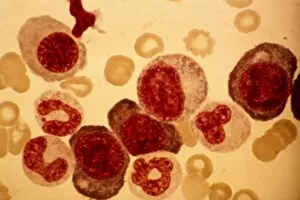
Background imageErythrocyte Collection: Pernicious anaemia, light micrograph

Pernicious anaemia, light micrograph
Pernicious anaemia. Light micrograph of megaloblast blood cells from bone marrow in a case of pernicious anaemia, also known as Biermers anaemia

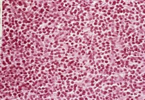
Background imageErythrocyte Collection: Blood cell cancer, light micrograph

Blood cell cancer, light micrograph
Blood cell cancer. Light micrograph of blood cells from a lymphatic ganglion in a case of a blood cancer of a mixed cell type

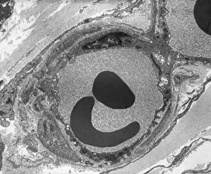
Background imageErythrocyte Collection: Capillary, TEM

Capillary, TEM
Capillary. Transmission electron micrograph (TEM) of a section through a capillary, showing two red blood cells (erythrocytes, black) in its interior

Background imageErythrocyte Collection: Blood cells, light micrograph

Blood cells, light micrograph
Blood cells. Light micrograph of red blood cells (erythrocytes, light blue) and white blood cells (leucocytes, nuclei stained purple)

Background imageErythrocyte Collection: Leukaemia cell, SEM

Leukaemia cell, SEM
Leukaemia cell. Coloured scanning electron micrograph (SEM) of a leukaemic (cancerous) lymphocyte white blood cell (green), amongst normal red blood cells (erythrocytes, red)

Background imageErythrocyte Collection: Reticulosarcoma, light micrograph

Reticulosarcoma, light micrograph
Reticulosarcoma. Light micrograph of reticulocyte blood cells (red, one at upper left) from a lymphatic ganglion in a case of reticulosarcoma

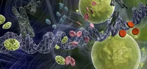
Background imageErythrocyte Collection: Drug effect on viruses, conceptual image C016 / 6253

Drug effect on viruses, conceptual image C016 / 6253
Drug effect on viruses, conceptual image. Computer artwork showing a single strand of DNA (deoxyribonucleic acid, spiral, centre), red blood cells (pink), virus particles (virions, green, small)

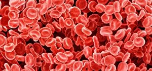
Background imageErythrocyte Collection: Red blood cells, SEM C015 / 8789

Red blood cells, SEM C015 / 8789
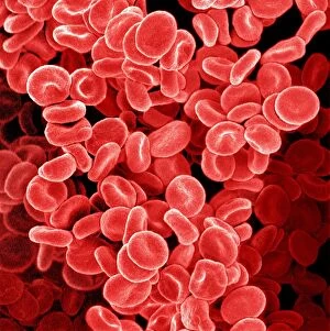
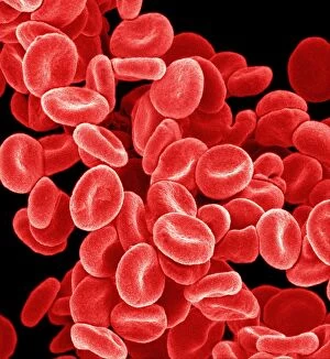
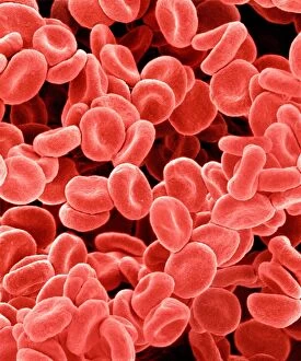
Red blood cells. Coloured scanning electron micrograph (SEM) of human red blood cells (erythrocytes). Red blood cells are biconcave, giving them a large surface area for gas exchange

Background imageErythrocyte Collection: Red blood cells, SEM C015 / 8792

Red blood cells, SEM C015 / 8792
Red blood cells. Coloured scanning electron micrograph (SEM) of human red blood cells (erythrocytes). Red blood cells are biconcave, giving them a large surface area for gas exchange

Background imageErythrocyte Collection: Red blood cells, SEM C015 / 8794

Red blood cells, SEM C015 / 8794
Red blood cells. Coloured scanning electron micrograph (SEM) of human red blood cells (erythrocytes). Red blood cells are biconcave, giving them a large surface area for gas exchange

Background imageErythrocyte Collection: Red blood cells, SEM C015 / 8796

Red blood cells, SEM C015 / 8796
Red blood cells. Coloured scanning electron micrograph (SEM) of human red blood cells (erythrocytes). Red blood cells are biconcave, giving them a large surface area for gas exchange

Background imageErythrocyte Collection: Red blood cells, SEM C015 / 8790

Red blood cells, SEM C015 / 8790
Red blood cells. Coloured scanning electron micrograph (SEM) of human red blood cells (erythrocytes). Red blood cells are biconcave, giving them a large surface area for gas exchange

Background imageErythrocyte Collection: Blood clot, SEM C015 / 9608

Blood clot, SEM C015 / 9608
Blood clot. Coloured scanning electron micrograph (SEM) of a blood clot from the inner wall of the left ventricle of a heart

Background imageErythrocyte Collection: Reticulosarcoma, light micrograph C015 / 7133

Reticulosarcoma, light micrograph C015 / 7133
Reticulosarcoma. Light micrograph of reticulocyte blood cells (red, one at upper left) from a lymphatic ganglion in a case of reticulosarcoma

Background imageErythrocyte Collection: Red blood cells, SEM C015 / 8793

Red blood cells, SEM C015 / 8793
Red blood cells. Coloured scanning electron micrograph (SEM) of human red blood cells (erythrocytes). Red blood cells are biconcave, giving them a large surface area for gas exchange

Background imageErythrocyte Collection: Red blood cells, SEM C015 / 8795

Red blood cells, SEM C015 / 8795
Red blood cells. Coloured scanning electron micrograph (SEM) of human red blood cells (erythrocytes). Red blood cells are biconcave, giving them a large surface area for gas exchange

Background imageErythrocyte Collection: Red blood cells, SEM C015 / 8787

Red blood cells, SEM C015 / 8787
Red blood cells. Coloured scanning electron micrograph (SEM) of human red blood cells (erythrocytes). Red blood cells are biconcave, giving them a large surface area for gas exchange


All Professionally Made to Order for Quick Shipping

"Erythrocyte: The Lifeline of Blood" Erythrocytes, commonly known as red blood cells, play a crucial role in our circulatory system. These tiny, disc-shaped cells are responsible for transporting oxygen from the lungs to every tissue and organ in our body. During menstruation, the uterus lining sheds and erythrocytes come into action. They help deliver necessary nutrients and oxygen to support the regrowth of this lining, ensuring a healthy reproductive cycle. Under scanning electron microscopy (SEM), these remarkable blood cells reveal their intricate structure. Their biconcave shape allows for maximum surface area exposure, facilitating efficient gas exchange within our bodies. The importance of erythrocytes extends beyond just oxygen transport. They also contribute to the complex process of blood coagulation cascade – an essential mechanism that prevents excessive bleeding when injuries occur. Artwork C016/9873 beautifully illustrates this intricate cascade that leads to clot formation. In SEM image C016/9747, we witness a close-up view of a blood clot formed by platelets and fibrin strands working together harmoniously to seal wounds effectively. Human red blood corpuscles captured under SEM showcase their vibrant nature while reminding us of their vital function within our bloodstream - maintaining homeostasis by carrying carbon dioxide back to the lungs for elimination. Even parasites like mouse malaria can be observed through SEM attached to erythrocytes - highlighting how these cells serve as hosts during infection but also become targets for immune responses against such invaders. Blood vessels act as highways where erythrocytes travel tirelessly throughout our bodies. These microscopic highways ensure proper distribution of nutrients and removal of waste products from various tissues they reach. SEM images further unveil intriguing details about erythrocyte behavior; one such example is seen in an image depicting a blood clot forming on plaster's surface – showcasing how these incredible cells respond swiftly when faced with injury or damage outside the body too.