Fraud Blocker Skip to main content

Eukaryotic Collection (page 2)

Eukaryotic organisms encompass a vast array of life forms, ranging from the microscopic to the towering kelp forests in our oceans

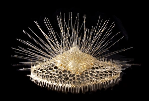
Background imageEukaryotic Collection: Eucyrtidium cranoides, radiolarian

Eucyrtidium cranoides, radiolarian
A glass model of a radiolarian, created by Leopold and Rudolf Blaschka in the late nineteenth century and held at the Natural History Museum, London

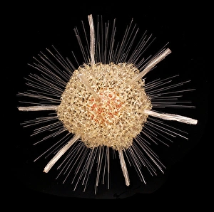
Background imageEukaryotic Collection: Actinophrys sol, heliozoan

Actinophrys sol, heliozoan
A glass model of a radiolarian, created by Leopold and Rudolf Blaschka in the late nineteenth century and held at the Natural History Museum, London

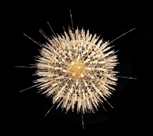
Background imageEukaryotic Collection: Heliosphaera actinota, radiolarian

Heliosphaera actinota, radiolarian
A glass model of a radiolarian, created by Leopold and Rudolf Blaschka in the late nineteenth century and held at the Natural History Museum, London

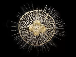
Background imageEukaryotic Collection: Eucecryphalus schultzei, radiolarian

Eucecryphalus schultzei, radiolarian
A glass model of a radiolarian, created by Leopold and Rudolf Blaschka in the late nineteenth century and held at the Natural History Museum, London

Background imageEukaryotic Collection: Spongosphaera streptacantha, radiolarian

Spongosphaera streptacantha, radiolarian
A glass model of a radiolarian, created by Leopold and Rudolf Blaschka in the late nineteenth century and held at the Natural History Museum, London

Background imageEukaryotic Collection: Aulosphaera elegantissima, radiolarian

Aulosphaera elegantissima, radiolarian
A glass model of a radiolarian, created by Leopold and Rudolf Blaschka in the late nineteenth century and held at the Natural History Museum, London

Background imageEukaryotic Collection: Difflugia pyriformis, amoebae

Difflugia pyriformis, amoebae
A glass model of amoebae, created by Leopold and Rudolf Blaschka in the late nineteenth century and held at the Natural History Museum, London

Background imageEukaryotic Collection: Radiolarian

Radiolarian
A glass model of a radiolarian, created by Leopold and Rudolf Blaschka in the late nineteenth century and held at the Natural History Museum, London

Background imageEukaryotic Collection: Syracosphaera anthos

Syracosphaera anthos
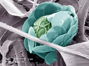
Coccosphere from the Western Mediterranean. False coloured to show the shell is formed of inner and outer layers of coccoliths with very different structure

Background imageEukaryotic Collection: Picture No. 11675491

Picture No. 11675491
Light Micrograph: Ciliate. Date:

Background imageEukaryotic Collection: Picture No. 11675490

Picture No. 11675490
Light Micrograph (LM): Elongate Ciliate and Paramecium bursaria. Date:

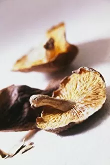
Background imageEukaryotic Collection: Dried shiitake mushrooms, defocused

Dried shiitake mushrooms, defocused

Background imageEukaryotic Collection: Ubiquitin activating enzyme protein E1 F007 / 9919

Ubiquitin activating enzyme protein E1 F007 / 9919
Molecular model of a ubiquitin-activating enzyme, also known as E1 enzymes. These catalyse the first step in the ubiquitination reaction, which targets a protein for degradation via a proteasome

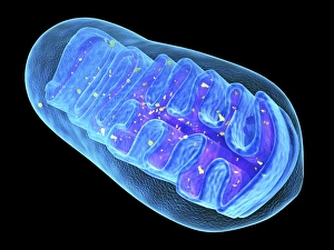
Background imageEukaryotic Collection: Mitochondria, artwork

Mitochondria, artwork
Mitochondria, computer artwork

Background imageEukaryotic Collection: Ubiquitin molecule F006 / 9528

Ubiquitin molecule F006 / 9528
Ubiquitin, molecular model. Ubiquitin is found in all eukaryotic cells. When a protein is damaged or old it will be tagged by several ubiquitin molecules
Background imageEukaryotic Collection: Golgi apparatus, artwork F006 / 9211

Golgi apparatus, artwork F006 / 9211
Computer artwork of the Golgi apparatus of the human cell. This organelle functions as a central delivery system for the cell

Background imageEukaryotic Collection: Mitochondrial structure, artwork F006 / 9207

Mitochondrial structure, artwork F006 / 9207
Mitochondrial structure. Computer artwork of a mitochondrion, showing a loop of mitochondrial DNA (deoxyribonucleic acid, mtDNA), the organelles genetic material

Background imageEukaryotic Collection: Golgi apparatus, artwork F006 / 9199

Golgi apparatus, artwork F006 / 9199
Computer artwork of the Golgi apparatus of the human cell. This organelle functions as a central delivery system for the cell

Background imageEukaryotic Collection: Mitochondrial structure, artwork F006 / 9198

Mitochondrial structure, artwork F006 / 9198
Mitochondrial structure. Computer artwork of a mitochondrion, showing a loop of mitochondrial DNA (deoxyribonucleic acid, mtDNA), the organelles genetic material

Background imageEukaryotic Collection: Golgi apparatus, artwork F006 / 9189

Golgi apparatus, artwork F006 / 9189
Computer artwork of the Golgi apparatus of the human cell. This organelle functions as a central delivery system for the cell

Background imageEukaryotic Collection: Cell membrane, artwork C018 / 7905

Cell membrane, artwork C018 / 7905
Cell membrane. Computer artwork of a section though a cell membrane. This is a semi-permeable membrane that controls what substances leave and enter the cell

Background imageEukaryotic Collection: Vault ribonucleoprotein molecule

Vault ribonucleoprotein molecule
Vault cytoplasmic ribonucleoprotein, molecular model. This molecule is made up from proteins and RNA (ribonucleic acid) and has a 39-fold symmetry

Background imageEukaryotic Collection: Kidney mitochondria, TEM

Kidney mitochondria, TEM
Kidney mitochondria. Transmission electron micrograph (TEM) of a section through a kidney tubule, showing numerous mitochondria (blue)

Background imageEukaryotic Collection: Eukaryote cell, artwork C016 / 6260

Eukaryote cell, artwork C016 / 6260
Eukaryote cell. Computer artwork showing the internal structure of a typical eukaryotic cell. Eukaryotes are organisms whose cells contain a membrane-bound nucleus (karyon, orange)

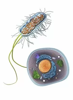
Background imageEukaryotic Collection: Prokaryote and eukaryote cells, artwork C016 / 6259

Prokaryote and eukaryote cells, artwork C016 / 6259
Prokaryote and eukaryote cells. Computer artwork comparing typical prokaryotic (top) and eukaryotic (bottom) cells. Prokaryotes are organisms whose cells lack a membrane-bound nucleus (karyon)

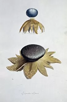
Background imageEukaryotic Collection: Fungus (Lycoperdon coliforme) C016 / 5988

Fungus (Lycoperdon coliforme) C016 / 5988
Fungus (Lycoperdon coliforme). Plate 1 from Drawings of Submerged Algae (1800) by Mary Dawson Turner. Held in the Botany Library at the Natural History Museum, London, UK

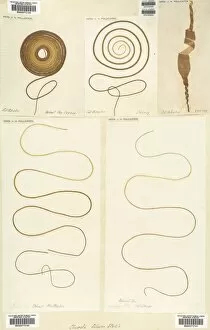
Background imageEukaryotic Collection: Dried seaweed specimens C016 / 5977

Dried seaweed specimens C016 / 5977
Dried seaweed (Chorda filum) specimens. Herbarium sheet from the collection held at the Natural History Museum, London, UK

Background imageEukaryotic Collection: Animal cell, illustration C018 / 0734

Animal cell, illustration C018 / 0734
Animal cell. Illustration showing the organelles present in an animal cell. At lower centre is the nucleus, which contains the cells genetic information in the form of DNA (deoxyribonucleic acid)

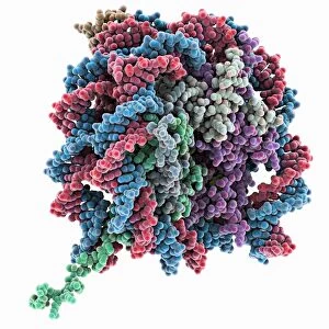
Background imageEukaryotic Collection: Nucleosome core particle bound to DNA C014 / 0872

Nucleosome core particle bound to DNA C014 / 0872
Nucleosome core particle bound to DNA. Molecular model showing a nucleosome core particle (green and purple) bound to a strand of DNA (deoxyribonucleic acid, blue and red)

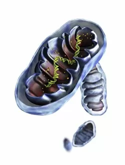
Background imageEukaryotic Collection: Mitochondrial structure, artwork

Mitochondrial structure, artwork
Mitochondrial structure. Computer artwork of a section through a mitochondrion, showing the internal structure and a loop of mitochondrial DNA (deoxyribonucleic acid, mtDNA)

Background imageEukaryotic Collection: Seaweed (Desmarestia ligulata) C016 / 6014

Seaweed (Desmarestia ligulata) C016 / 6014
seaweed (Desmarestia ligulata). Page 55 from Algae Danmonienses: or dried specimens of Marine Plants, principally collected in Devonshire by Mary Wyatt; carefully named according to Dr

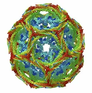
Background imageEukaryotic Collection: Clathrin lattice, molecular model C015 / 6788

Clathrin lattice, molecular model C015 / 6788
Molecular model of a complete clathrin lattice. The polyhedral protein lattice coats eukaryotic cell membranes (vesicles) and coated pits and appears to be involved in protein secretion

Background imageEukaryotic Collection: Clathrin lattice, molecular model C015 / 6790

Clathrin lattice, molecular model C015 / 6790
Molecular model of a complete clathrin lattice. The polyhedral protein lattice coats eukaryotic cell membranes (vesicles) and coated pits and appears to be involved in protein secretion

Background imageEukaryotic Collection: Mitochondrial structure, artwork C015 / 6784

Mitochondrial structure, artwork C015 / 6784
Mitochondrial structure. Computer artwork of a mitochondrion, showing a loop of mitochondrial DNA (deoxyribonucleic acid, mtDNA), the organelles genetic material

Background imageEukaryotic Collection: Clathrin lattice, molecular model C015 / 6770

Clathrin lattice, molecular model C015 / 6770
Molecular model of a complete clathrin lattice. The polyhedral protein lattice coats eukaryotic cell membranes (vesicles) and coated pits and appears to be involved in protein secretion

Background imageEukaryotic Collection: Golgi apparatus, artwork C015 / 6764

Golgi apparatus, artwork C015 / 6764
Computer artwork of the Golgi apparatus of the human cell. This organelle functions as a central delivery system for the cell

Background imageEukaryotic Collection: Mitochondrial structure, artwork C015 / 6765

Mitochondrial structure, artwork C015 / 6765
Mitochondrial structure. Computer artwork of a mitochondrion, showing a loop of mitochondrial DNA (deoxyribonucleic acid, mtDNA), the organelles genetic material

Background imageEukaryotic Collection: Carrageen moss (Chondus cripus) C016 / 6062

Carrageen moss (Chondus cripus) C016 / 6062
Carrageen moss (Chondus cripus). Specimen from Algae Danmonienses: or dried specimens of Marine Plants, principally collected in Devonshire by Mary Wyatt; carefully named according to Dr

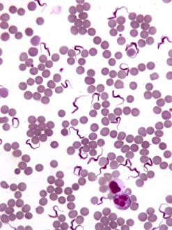
Background imageEukaryotic Collection: Trypanosomes in blood smear, SEM C016 / 5783

Trypanosomes in blood smear, SEM C016 / 5783
Parasitic protozoan. Coloured scanning electron micrograph (SEM) of trypanosome protozoa (Trypanosoma sp.) in a blood smear

Background imageEukaryotic Collection: Human cell, artwork C013 / 6522

Human cell, artwork C013 / 6522
Human cell. Illustration of a section through a human cell. At centre is the nucleus (purple), with the nucleolus (dark red) inside it

Background imageEukaryotic Collection: Dinoflagellate protozoan, SEM C013 / 5111

Dinoflagellate protozoan, SEM C013 / 5111
Dinoflagellate protozoan, scanning electron micrograph (SEM). Dinoflagellates are unicellular protozoans. About 90 percent are found in marine environments as plankton

Background imageEukaryotic Collection: Coccolithophorid plankton, SEM C013 / 5109

Coccolithophorid plankton, SEM C013 / 5109
Coccolithophorid plankton. Coloured scanning electron micrograph (SEM) of the external mineralised structure (coccosphere) of a small marine algal organism called a coccolithophore

Background imageEukaryotic Collection: Heron- Allen microscope slides of foraminifera

Heron- Allen microscope slides of foraminifera
Christmas greetings spelt out in minute shells which bears the initials of the maker, Edward Heron-Allen, and the year it was made: E H A, 1909


All Professionally Made to Order for Quick Shipping

Eukaryotic organisms encompass a vast array of life forms, ranging from the microscopic to the towering kelp forests in our oceans. Take a closer look at these captivating hints that showcase the diversity and complexity of eukaryotes. Starting with budding yeast cells, we witness their remarkable ability to reproduce through cell division. Under the scanning electron microscope (SEM), dividing yeast cells reveal intricate structures and processes that contribute to their survival and proliferation. Moving on to pressed seaweed specimens, such as Dictyota dichotoma and Fucus bulbosus, we explore the fascinating world of marine algae. These eukaryotic organisms play crucial roles in ocean ecosystems by providing shelter for countless other species while also contributing to nutrient cycling. Delving deeper into microscopic wonders, we encounter Diatoms under SEM. These single-celled eukaryotes exhibit stunning geometric patterns on their silica-based shells. Their ecological significance cannot be overstated as they are responsible for a significant portion of Earth's oxygen production. Another mesmerizing microorganism is Discosphaera tubifera, commonly known as coccolithophore. These tiny calcifying algae adorn themselves with intricately sculpted calcium carbonate plates called coccoliths. Their presence can create breathtaking blooms visible from space. Shifting gears towards Plasmodium sp. , an insidious malarial parasite that infects human red blood cells, we confront one of the darker aspects lifeforms. This reminder highlights how even within this kingdom there exists both beauty and danger. To round out our exploration, let's not forget about majestic kelps like Fucus radiatus. These large brown algae form underwater forests teeming with biodiversity while serving as vital carbon sinks in our changing climate. Finally, artistic renditions depicting various cell types remind us that behind every scientific discovery lies creativity and imagination – essential tools for unraveling nature's mysteries.