Fraud Blocker Skip to main content

Excretory Collection

"Exploring the Intricacies of Excretory System

Background imageExcretory Collection: Kidney glomeruli, SEM

Kidney glomeruli, SEM
Kidney glomeruli, coloured scanning electron micrograph (SEM). The cells that usually surround the glomeruli have been removed to reveal the coiled capillaries at the core of the glomeruli

Background imageExcretory Collection: Kidney glomerulus, SEM

Kidney glomerulus, SEM
Kidney glomerulus, coloured scanning electron micrograph (SEM). Podocyte cells, which make up the surface of the glomerulus, are yellow. Magnification: x650 when printed at 10 centimetres wide

Background imageExcretory Collection: Fibroblast cell, artwork

Fibroblast cell, artwork
Fibroblast cell. Computer artwork of a fibroblast excreting collagen fibres (tropocollagen). Fibroblasts are cells that produce connective tissue such as collagen (tropocollagen)

Background imageExcretory Collection: Kidneys, anatomical artwork

Kidneys, anatomical artwork
Kidneys. Anatomical computer artwork of a mans body, showing the position of the kidneys (red, centre left and right) and adrenal glands (beige)

Background imageExcretory Collection: Renal blood supply, anatomical artwork

Renal blood supply, anatomical artwork
Renal blood supply. Anatomical computer artwork of a human kidney (left) and adrenal gland (beige, upper centre), showing their main blood supply

Background imageExcretory Collection: Kidney anatomy, artwork C016 / 8945

Kidney anatomy, artwork C016 / 8945
Kidney anatomy, artwork. The kidneys filter waste from the blood and excrete it as urine. The kidney consists of an outer cortex and an inner medulla

Background imageExcretory Collection: Human kidneys, historical model

Human kidneys, historical model
Human kidneys. 1950s anatomical model of the human kidneys (red) with the ureters (tubes, upper left) attached, and a section through one kidney showing the internal structure

Background imageExcretory Collection: Kidney anatomy, artwork C016 / 8663

Kidney anatomy, artwork C016 / 8663
Kidney anatomy, artwork. The kidneys filter waste from the blood and excrete it as urine. The kidney consists of an outer cortex and an inner medulla

Background imageExcretory Collection: Kidney anatomy, artwork C016 / 8661

Kidney anatomy, artwork C016 / 8661
Kidney anatomy, artwork. The kidneys filter waste from the blood and excrete it as urine. The kidney consists of an outer cortex and an inner medulla

Background imageExcretory Collection: Kidney anatomy, artwork C016 / 8662

Kidney anatomy, artwork C016 / 8662
Kidney anatomy, artwork. The kidneys filter waste from the blood and excrete it as urine. The kidney consists of an outer cortex and an inner medulla

Background imageExcretory Collection: Kidney anatomy, artwork C016 / 8660

Kidney anatomy, artwork C016 / 8660
Kidney anatomy, artwork. The kidneys filter waste from the blood and excrete it as urine. The kidney consists of an outer cortex and an inner medulla

Background imageExcretory Collection: Urinary system, artwork C015 / 6455

Urinary system, artwork C015 / 6455
Urinary system. Computer artwork showing the organs of the human urinary system. The kidneys (red, top left and right) filter waste products such as urea from the blood (red and blue) to form urine

Background imageExcretory Collection: Urinary system, artwork C015 / 6454

Urinary system, artwork C015 / 6454
Urinary system. Computer artwork showing the organs of the human urinary system. The kidneys (red, top left and right) filter waste products such as urea from the blood to form urine

Background imageExcretory Collection: Kidney glomerulus, TEM C016 / 5831

Kidney glomerulus, TEM C016 / 5831
Kidney glomerulus. Transmission electron micrograph (TEM) of a section through a renal glomerulus. At centre left is a capillary with a red blood cell (black) in its lumen

Background imageExcretory Collection: Penicillin research, World War II C016 / 4300

Penicillin research, World War II C016 / 4300
Penicillin research. Graph showing the rate of excretion of the antibiotic drug penicillin following intravenous administration. The horizontal axis shows time in hours

Background imageExcretory Collection: Sweat gland in skin

Sweat gland in skin. Light micrograph of a section through the dermis of the skin showing a sweat gland. The gland is composed of a secretory portion (upper left)

Background imageExcretory Collection: Renal blood vessels, X-ray

Renal blood vessels, X-ray
Renal blood vessels. Angiogram (X-ray) of blood vessels in a healthy kidney. The large artery entering the kidney is the renal artery, which branches into many smaller capillaries inside the kidney

Background imageExcretory Collection: Kidney podocyte cells, SEM

Kidney podocyte cells, SEM
Kidney podocyte cells. Coloured scanning electron micrograph (SEM) of podocyte cells on the surface of a kidney glomerulus. Podocytes possess branching, foot-like processes (red/brown)

Background imageExcretory Collection: Kidney filtration, SEM

Kidney filtration, SEM
Kidney filtration. Coloured scanning electron micrograph (SEM) of tissue from a healthy kidney, showing the glomerular structure. The surface of a glomerulus is encased in podocyte cells (purple)

Background imageExcretory Collection: Pancreas cells, SEM

Pancreas cells, SEM
Pancreas cells. Coloured scanning electron micrograph (SEM) of acinar (exocrine) pancreatic cells. Acinar cells produce and excrete digestive enzymes to the small intestine, via the pancreatic ducts

Background imageExcretory Collection: Kidneys, abdominal CT scan

Kidneys, abdominal CT scan
Normal kidneys. Coloured axial (horizontal) CT (computed tomography) scan through a human abdomen at the level of the kidneys. The front of the body is at top

Background imageExcretory Collection: Abdominal bones, 3-D CT scan

Abdominal bones, 3-D CT scan
Abdominal bones, coloured 3-D computer tomography (CT) scan. The lower part of the ribcage is in upper frame, with the pelvis in lower frame. They are connected by the spine, which runs down centre

Background imageExcretory Collection: Kidney glomeruli, light micrograph

Kidney glomeruli, light micrograph
Kidney glomeruli. Light microscope of a section through the cortex of a healthy kidney showing glomeruli. The core of a glomerulus is formed of a tightly coiled capillary (purple)

Background imageExcretory Collection: Human urinary system, artwork

Human urinary system, artwork
Urinary system. Computer artwork of a male torso and the urinary system, showing kidney, urethra, bladder and suprarenal gland

Background imageExcretory Collection: Pancreas cell, SEM

Pancreas cell, SEM
Pancreas cell. Coloured scanning electron micrograph (SEM) of an acinar (exocrine) pancreatic cell. Acinar cells produce and excrete digestive enzymes to the small intestine

Background imageExcretory Collection: Pancreas tissue, SEM

Pancreas tissue, SEM
Pancreas tissue. Coloured scanning electron micrograph (SEM) of fractured pancreas tissue, showing numerous acinar cells, containing secretory zymogen granules

Background imageExcretory Collection: Glomerular capillary, TEM

Glomerular capillary, TEM
Glomerular capillary. Coloured transmission electron micrograph (TEM) of a section through kidney tissue, showing a capillary from a glomerulus. Glomeruli filter waste products from the blood

Background imageExcretory Collection: Kidney glomerulus surface, SEM

Kidney glomerulus surface, SEM
Kidney glomerulus surface. Coloured scanning electron micrograph (SEM) of podocyte cells (orange) on the surface of a kidney glomerulus. These have branching processes (pedicles) that interlock

Background imageExcretory Collection: Kidney glomerulus, light micrograph

Kidney glomerulus, light micrograph
Kidney glomerulus. Coloured light micrograph of a section through the cortex of a healthy kidney, showing a glomerulus. The core of a glomerulus is formed of a tightly coiled capillary (purple)

Background imageExcretory Collection: Female urinary system, artwork

Female urinary system, artwork
Female urinary system. Computer artwork of a naked woman with a healthy urinary system. The kidneys can be seen (upper left and right), as well as the ureters

Background imageExcretory Collection: Alcohol Advertising 1896

Alcohol Advertising 1896
Aperal - aperitif which may be drunk on its own or mixed with your coffee; either way it excites your appetite & benefits your excretory organs


All Professionally Made to Order for Quick Shipping

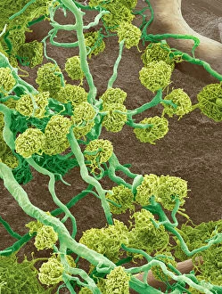
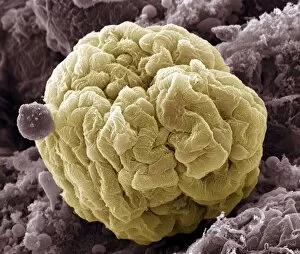
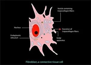
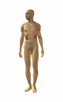
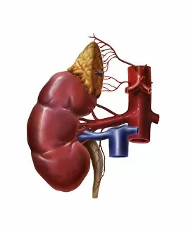
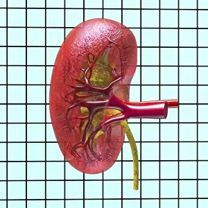
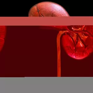
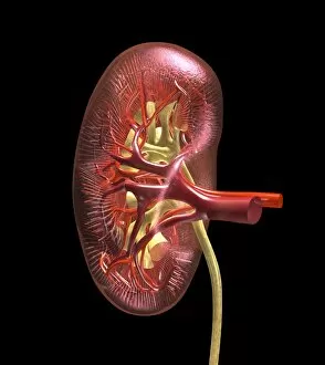
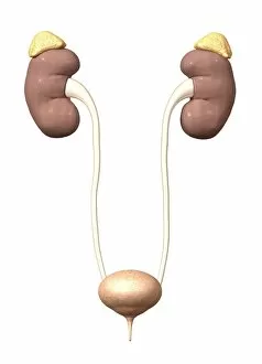
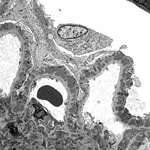
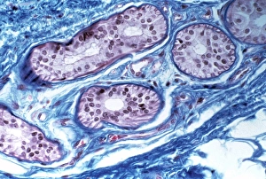
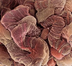
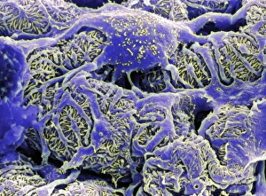
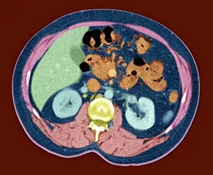
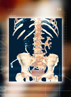
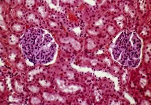
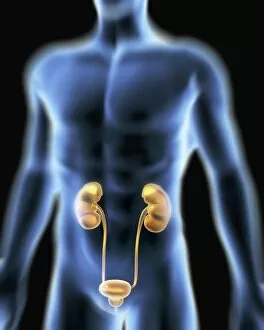
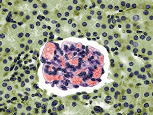
"Exploring the Intricacies of Excretory System: A Journey through Kidney Glomeruli and Beyond" Delve into the fascinating world of excretion as we embark on a microscopic adventure. Witness the intricate beauty of kidney glomerulus, captured in stunning detail by scanning electron microscopy (SEM). These tiny structures, resembling delicate webs, play a crucial role in filtering waste from our blood. Marvel at the complexity of kidney glomeruli, as SEM unveils their intricate network. These remarkable filters ensure that only essential substances are retained while waste products are efficiently eliminated. The interconnectedness of these glomeruli is truly awe-inspiring. Zooming out to explore further, SEM reveals the intricacy of kidney blood vessels. Like lifelines coursing through our bodies, they transport vital nutrients and oxygen to keep our kidneys functioning optimally. Switching gears from scientific imagery to artistic representation, an exquisite artwork portrays fibroblast cells within our kidneys. These specialized cells provide structural support and contribute to tissue repair when needed. Returning to SEM's captivating lens once again showcases multiple kidney glomeruli in all their glory. Their unique shapes and arrangements emphasize their importance in maintaining proper renal function. A light micrograph offers another perspective on kidney glomeruli – this time with enhanced contrast and clarity under different imaging techniques. This glimpse into their inner workings highlights how each component contributes harmoniously towards efficient filtration. Shifting focus momentarily from kidneys alone, an artwork depicts a healthy digestive system - reminding us that excretion is just one part of a larger bodily process ensuring overall well-being. Revisiting anatomical artworks dedicated solely to kidneys emphasizes their significance within our bodies' framework. They serve as silent heroes tirelessly regulating fluid balance, electrolyte levels, and removing waste products day after day. Anatomical artworks showcasing renal blood supply take us on yet another visual journey highlighting how these organs receive nourishment for optimal functionality - underscoring the importance of a healthy circulatory system.