Fraud Blocker Skip to main content

Fasciculus Collection

"Fasciculus: Exploring the Intricate Connections of Flanders" Step into the world of Fasciculus

Background imageFasciculus Collection: Coloured SEM of a section through nerve fibres

Coloured SEM of a section through nerve fibres
Nerve fibres. Coloured scanning electron micrograph (SEM) of a section through bundles of nerve fibres (red). Each is within a sheath, or perineurium (blue)

Background imageFasciculus Collection: Map Of Flanders By Cartographer Matthias Quad From His Fasciculus Geographicus. Later Hand Colouring

Map Of Flanders By Cartographer Matthias Quad From His Fasciculus Geographicus. Later Hand Colouring. Engraved And Published By Johannes Bussemacher, Cologne, 1608

Background imageFasciculus Collection: Brains white matter, DTI MRI scan

Brains white matter, DTI MRI scan
Brains white matter. Close-up of an area of the brain imaged using tract density imaging and 3D diffusion tensor imaging (DTI), a magnetic resonance imaging (MRI) technique

Background imageFasciculus Collection: Human muscular structure, artwork F008 / 0250

Human muscular structure, artwork F008 / 0250
Human muscular structure, computer artwork

Background imageFasciculus Collection: Human muscular structure, artwork F008 / 0443

Human muscular structure, artwork F008 / 0443
Human muscular structure, computer artwork

Background imageFasciculus Collection: Human muscular structure, artwork F008 / 0167

Human muscular structure, artwork F008 / 0167
Human muscular structure, computer artwork

Background imageFasciculus Collection: Human muscular structure, artwork F008 / 1021

Human muscular structure, artwork F008 / 1021
Human muscular structure, computer artwork

Background imageFasciculus Collection: Human muscular structure, artwork F008 / 0143

Human muscular structure, artwork F008 / 0143
Human muscular structure, computer artwork

Background imageFasciculus Collection: Human muscular structure, artwork F008 / 0442

Human muscular structure, artwork F008 / 0442
Human muscular structure, computer artwork

Background imageFasciculus Collection: Human muscular structure, artwork F008 / 0249

Human muscular structure, artwork F008 / 0249
Human muscular structure, computer artwork

Background imageFasciculus Collection: Human muscular structure, artwork F008 / 1202

Human muscular structure, artwork F008 / 1202
Human muscular structure, computer artwork

Background imageFasciculus Collection: Human muscular structure, artwork F008 / 0317

Human muscular structure, artwork F008 / 0317
Human muscular structure, computer artwork

Background imageFasciculus Collection: Human muscular structure, artwork F008 / 1019

Human muscular structure, artwork F008 / 1019
Human muscular structure, computer artwork

Background imageFasciculus Collection: Human muscular structure, artwork F008 / 1200

Human muscular structure, artwork F008 / 1200
Human muscular structure, computer artwork

Background imageFasciculus Collection: Female reproductive system, artwork F008 / 0441

Female reproductive system, artwork F008 / 0441
Female reproductive system, computer artwork

Background imageFasciculus Collection: Human muscular structure, artwork F008 / 1022

Human muscular structure, artwork F008 / 1022
Human muscular structure, computer artwork

Background imageFasciculus Collection: Human muscular structure, artwork F008 / 0449

Human muscular structure, artwork F008 / 0449
Human muscular structure, computer artwork

Background imageFasciculus Collection: Human muscular structure, artwork F008 / 0164

Human muscular structure, artwork F008 / 0164
Human muscular structure, computer artwork

Background imageFasciculus Collection: Human muscular structure, artwork F008 / 1201

Human muscular structure, artwork F008 / 1201
Human muscular structure, computer artwork

Background imageFasciculus Collection: Human muscular structure, artwork F008 / 1204

Human muscular structure, artwork F008 / 1204
Human muscular structure, computer artwork

Background imageFasciculus Collection: Human muscular structure, artwork F008 / 0144

Human muscular structure, artwork F008 / 0144
Human muscular structure, computer artwork

Background imageFasciculus Collection: Atlanto-axial joint, artwork C016 / 6552

Atlanto-axial joint, artwork C016 / 6552
Atlanto-axial joint. Artwork of the bones (yellow) and ligaments (blue) of the atlanto-axial joint of the upper neck, as seen from above. The rear of the spine is at bottom

Background imageFasciculus Collection: KETHAM, Johannes de (second half 15th century-begin

KETHAM, Johannes de (second half 15th century-begin
" KETHAM, Johannes de (second half 15th century-begin 16th). Human health compendium" (Fasciculus medicinae, 1491). First medicine book including anatomical illustrations

Background imageFasciculus Collection: Nerve fibre, SEM

Nerve fibre, SEM
Nerve fibres. Coloured scanning electron micrograph (SEM) of nerve fibres (brown). A group of nerves such as this is known as a fasciculus

Background imageFasciculus Collection: Coloured SEM of some nerve fibres

Coloured SEM of some nerve fibres
Nerve fibres. Coloured scanning electron micrograph (SEM) of some nerve fibres. A group of nerves such as this is known as a fasciculus

Background imageFasciculus Collection: False-colour SEM of skeletal muscle fibres

False-colour SEM of skeletal muscle fibres
Muscle. False-colour scanning electron micrograph of a bundle of skeletal muscle fibres (red), also known as striated muscle fibres, and a few capillaries (blue)


All Professionally Made to Order for Quick Shipping

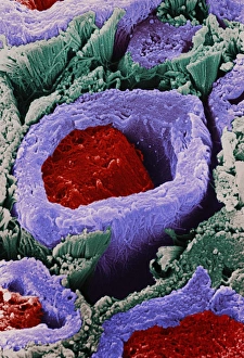
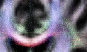
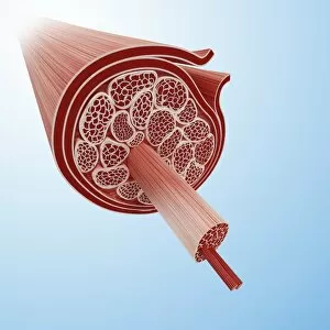
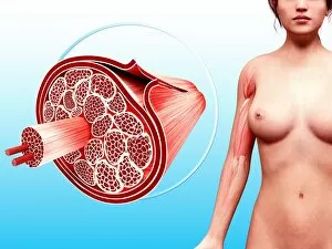
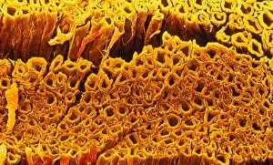
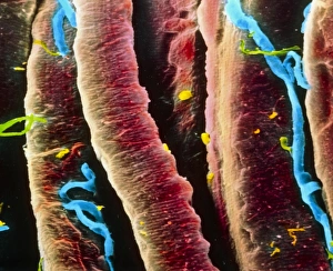
"Fasciculus: Exploring the Intricate Connections of Flanders" Step into the world of Fasciculus, a captivating journey through the historical and anatomical wonders of Flanders. This 1508 map by cartographer Matthias Quad, part of his renowned work "Fasciculus Geographicus, " takes us back in time to explore this enchanting region. With delicate hand coloring added later by Johannes Bussemacher, Cologne, this map showcases the intricate details and geographical features that make Flanders so unique. From its charming towns to its vast landscapes, every corner is meticulously depicted for your exploration. But our adventure doesn't stop there. Delve deeper into the human body's complex structure with Ketham's brain white matter DTI MRI scan. This groundbreaking technology allows us to visualize how nerve fibers interconnect within our brains, revealing a mesmerizing network responsible for our thoughts and actions. As we shift focus from maps to anatomy, let's marvel at the human muscular structure captured in stunning artwork. The various illustrations - F008/0250, F008/0443, F008/0167 - showcase muscles in all their glory. Each fiber intricately woven together forms an awe-inspiring system that enables movement and strength within our bodies. From biceps to quadriceps and everything in between, these artworks highlight both beauty and functionality. Whether it be flexing or extending muscles during physical activity or simply appreciating their form as works of art themselves; they remind us of the incredible capabilities residing within each one of us. So join us on this fascinating expedition through time and biology as we unravel the secrets hidden within Fasciculus Geographicus' pages. Discover not only the rich history etched onto maps but also marvel at nature's masterpiece –the human body– where every muscle tells a story waiting to be explored.