
Female Reproductive System Collection
The female reproductive system is a complex and intricate network of organs that play a crucial role in the creation of life


All Professionally Made to Order for Quick Shipping
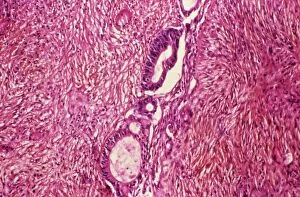
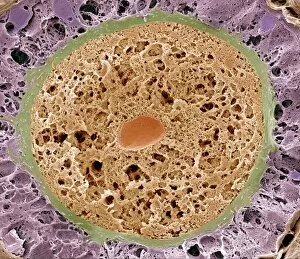
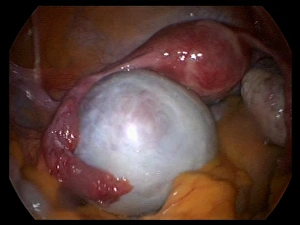
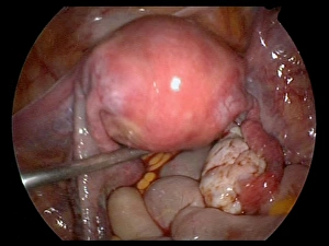
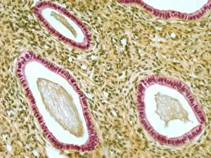
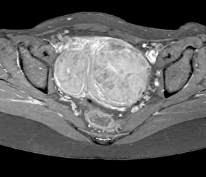
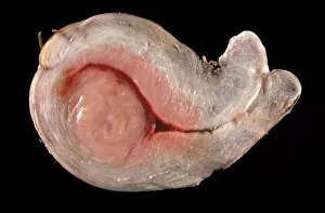
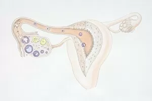
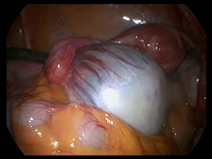
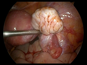
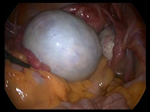
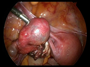
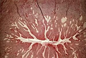
The female reproductive system is a complex and intricate network of organs that play a crucial role in the creation of life. From the ovaries to the uterus, each component serves its unique purpose in this miraculous process. Ovarian cancer, a devastating disease that affects countless women worldwide, reminds us of the importance of understanding and caring for our bodies. A light micrograph C015 / 7103 reveals the delicate nature of these vital organs, urging us to prioritize regular check-ups and screenings. Within the ovaries lie ovarian follicles, as seen through a scanning electron microscope (SEM). These tiny structures house immature eggs waiting for their chance at fertilization. The beauty captured by this image highlights both their fragility and potential. A cross-sectional view showcases an anterior perspective of the uterus with fibroids alongside other essential components like the vagina and cervix. This visual representation emphasizes how interconnected these parts are in supporting reproduction while also reminding us that conditions such as fibroids can impact fertility. An endoscope view C017 / 6800 allows us to witness an ovarian cyst firsthand. Although often benign, these fluid-filled sacs can cause discomfort or complications if left untreated. Regular medical examinations help detect them early on before they become problematic. Similarly, another endoscope view C017 / 6805 focuses on capturing an intimate glimpse into the uterus itself. By examining this organ closely, doctors can identify any abnormalities or signs of disease promptly—empowering women to take control over their reproductive health. Microscopic analysis provides further insight into specific components within this intricate system: a light micrograph F006 / 9805 offers a close-up look at cervical cells while another F006 / 9799 zooms in on fallopian tubes' inner workings—the very pathways where conception occurs. Uterine fibroids are common growths found within many women's uteruses; MRI scan C018 / 0466 helps visualize their size and location.