
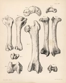
Femur Collection
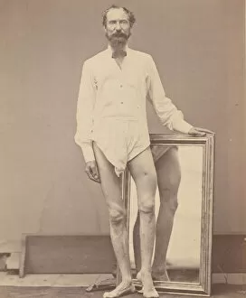
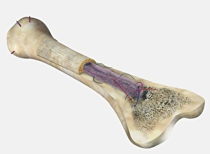
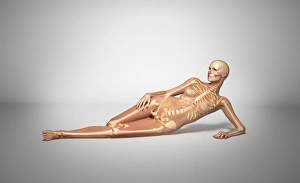
The femur, also known as the thigh bone, is a vital component of the human knee joint. It plays a crucial role in supporting our body weight and facilitating movement


All Professionally Made to Order for Quick Shipping
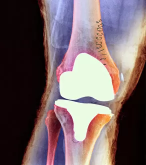
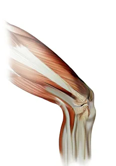
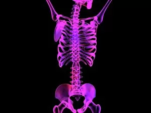
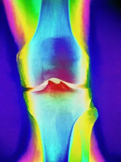
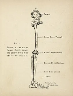
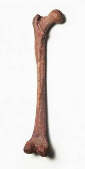
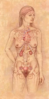
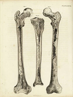
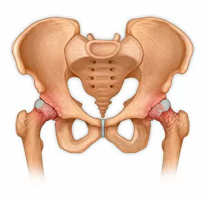
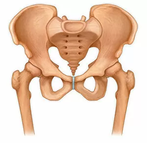
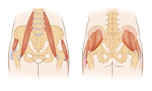
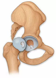
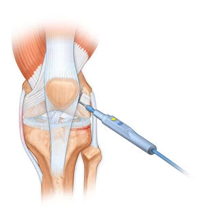
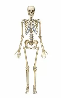
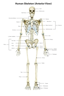
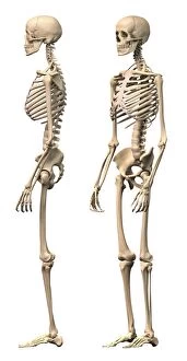
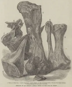
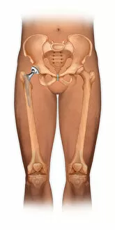
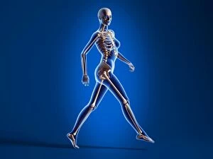
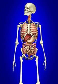
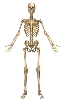
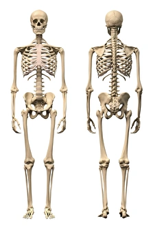
The femur, also known as the thigh bone, is a vital component of the human knee joint. It plays a crucial role in supporting our body weight and facilitating movement. From hip replacements to knee joint prostheses, medical advancements have revolutionized the treatment of femur-related conditions. An X-ray image reveals the intricate details of a total hip replacement, showcasing how this procedure can restore mobility and alleviate pain for individuals with damaged hips. Similarly, an artwork depicting a damaged knee ligament reminds us of the importance of maintaining strong and healthy joints. Colored X-rays provide fascinating insights into the complexity of a human knee joint. These images showcase its various components and highlight any potential issues that may require medical attention. Intriguing computer artwork showcases an upper body skeleton, emphasizing the significance of proper skeletal structure for overall health and well-being. A diagram illustrating the bones of the right leg and hip helps us visualize how these structures work together seamlessly to support our movements. A healthy knee is depicted in another X-ray image, serving as a reminder to take care of our joints through exercise and proper nutrition. However, cautionary symbols like inverted skull and crossbones warn against neglecting their maintenance or engaging in activities that could lead to injury. Lastly, we are reminded that even ancient history holds secrets about femurs - such as C016 / 5028 from Red Lady Paviland - highlighting both their enduring presence throughout time and their continued importance in understanding human anatomy.