
Fifty Two Collection
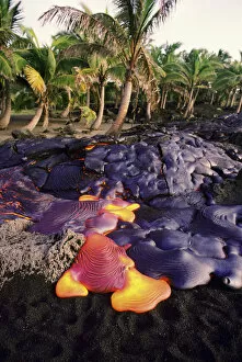
Exploring the wonders of Hawaii's Volcanoes National Park on Big Island


All Professionally Made to Order for Quick Shipping
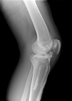
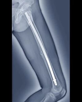
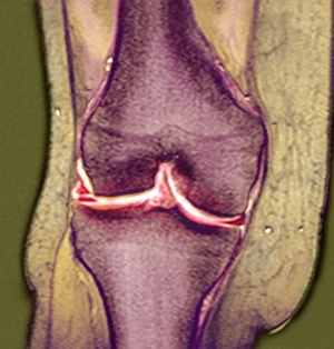
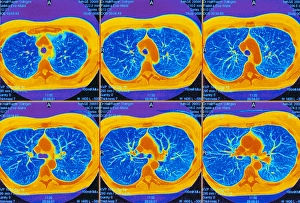
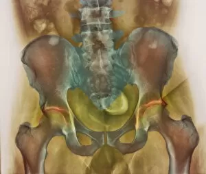
Exploring the wonders of Hawaii's Volcanoes National Park on Big Island, where the raw power of nature is on full display with the ongoing eruption of Kilauea in its phase 52. Meanwhile, back in the USA, medical files reveal X-rays of various diagnoses: melorheostosis of the knee (C017 / 7144, 7145, 7146), pinned thigh fractures (C016 / 6563, 6564), and knee meniscus tears. Elsewhere, CT scans offer a closer look into the intricacies of the lungs, while an osteoarthritic pelvis (as seen on X-rays) serves as a reminder of the wear and tear our bodies endure over time.