Fraud Blocker Skip to main content

Food Poisoning Collection

"Unseen Culprits: Exploring the Microscopic World of Food Poisoning" E

Background imageFood Poisoning Collection: E. coli bacteria, SEM

E. coli bacteria, SEM
E. coli bacteria. Coloured scanning electron micrograph (SEM) of Escherichia coli bacteria. These bacteria are a normal part of the intestinal flora in humans and other animals

Background imageFood Poisoning Collection: Salmonella bacteria, SEM

Salmonella bacteria, SEM
Salmonella bacteria, coloured scanning electron micrograph (SEM). These Gram-negative rod-shaped bacteria can cause food poisoning when eaten in contaminated food

Background imageFood Poisoning Collection: Norovirus particles, TEM

Norovirus particles, TEM
Norovirus particles. Coloured transmission electron micrograph (TEM) of norovirus particles. Norovirus is a genus of RNA (ribonucleic acid)

Background imageFood Poisoning Collection: E. coli bacteria

E. coli bacteria
False-colour transmission electron micrograph of the bacterium Escherichia coli, a normal inhabitant of the human intestine

Background imageFood Poisoning Collection: E. coli bacteria, SEM

E. coli bacteria, SEM
E. coli bacteria. Coloured scanning electron micrograph (SEM) of Escherichia coli bacteria (purple) taken from the small intestine of a child. E

Background imageFood Poisoning Collection: Salmonella bacteria, SEM

Salmonella bacteria, SEM
Salmonella bacteria, coloured scanning electron micrograph (SEM). Each rod-shaped structure is an individual bacterium. These bacteria occur mainly in human and animal intestines

Background imageFood Poisoning Collection: Salmonella bacteria, artwork C013 / 8818

Salmonella bacteria, artwork C013 / 8818
Salmonella bacteria, computer artwork. Salmonella sp. bacteria are gram-negative rod-shaped bacteria that have flagella (hair-like structures) that they use for locomotion

Background imageFood Poisoning Collection: Salmonella culture

Salmonella culture
MODEL RELEASED. Salmonella culture. Microbiologist holding a petri dish containing a culture of Salmonella sp. bacteria. These bacteria are the cause of salmonellosis (food poisoning) in humans

Background imageFood Poisoning Collection: E. coli 0157: H7 bacteria

E. coli 0157: H7 bacteria
E. coli 0157:H7 bacteria. Coloured scanning electron micrograph (SEM) of Escherichia coli 0157:H7 bacteria, cause of foodborne illness

Background imageFood Poisoning Collection: Salmonella bacterium dividing, SEM

Salmonella bacterium dividing, SEM
Salmonella bacterium dividing. Coloured scanning electron micrograph (SEM) of of a Salmonella bacterium dividing. The two new daughter cells are seen at upper right and lower left

Background imageFood Poisoning Collection: Cartoon depicting a man, suffering with food poisoning, asking the pharmacist for a cure

Cartoon depicting a man, suffering with food poisoning, asking the pharmacist for a cure, 19th century
5309344 Cartoon depicting a man, suffering with food poisoning, asking the pharmacist for a cure, 19th century; (add.info.: Cartoon depicting a man, suffering with food poisoning)

Background imageFood Poisoning Collection: Colony of Salmonella Typhi showing Bacilli with flagella, 1906 (litho)

Colony of Salmonella Typhi showing Bacilli with flagella, 1906 (litho)
3501916 Colony of Salmonella Typhi showing Bacilli with flagella, 1906 (litho) by German School, (20th century); Private Collection; (add.info)

Background imageFood Poisoning Collection: Colony of Salmonella typhi, 1906 (litho)

Colony of Salmonella typhi, 1906 (litho)
3501915 Colony of Salmonella typhi, 1906 (litho) by German School, (20th century); Private Collection; (add.info.: Bacteria constitute a large domain of prokaryotic microorganisms)

Background imageFood Poisoning Collection: Staphylococcus colony, 1905 (litho)

Staphylococcus colony, 1905 (litho)
3501895 Staphylococcus colony, 1905 (litho) by German School, (20th century); Private Collection; (add.info.: Bacteria constitute a large domain of prokaryotic microorganisms)

Background imageFood Poisoning Collection: Botulism bacteria

Botulism bacteria. Light micrograph of botulism bacteria (Clostridium botulinum). These bacteria occur naturally in soil. They produce botulinum toxin

Background imageFood Poisoning Collection: Clostridium perfringens bacterium with spore

Clostridium perfringens bacterium with spore
False-colour transmission electron micrograph (TEM) of a Clostridium perfringens bacterium with endospore. This species of Gram-positive

Background imageFood Poisoning Collection: Coloured SEM of Clostridium botulinum

Coloured SEM of Clostridium botulinum
Food poisoning bacterium. Coloured scanning electron micrograph of the Gram-positive anaerobic bacteria Clostridium botulinum, responsible for serious food poisoning in humans. C

Background imageFood Poisoning Collection: Conceptual image of salmonella typhi causing typhoid

Conceptual image of salmonella typhi causing typhoid

Background imageFood Poisoning Collection: Microscopic view of Listeria monocytogenes

Microscopic view of Listeria monocytogenes. Listeria monocytogenes is the causative agent of the bacterial infection called listeriosis

Background imageFood Poisoning Collection: Conceptual image of salmonella

Conceptual image of salmonella. Salmonella is a genus of rod-shaped, gram-negative bacteria. There are two main species of Salmonella, Salmonella bongori and Salmonella enterica

Background imageFood Poisoning Collection: Sirloin Steak

Sirloin Steak

Background imageFood Poisoning Collection: Two Pink Shrimp (Malacostracans), close up

Two Pink Shrimp (Malacostracans), close up

Background imageFood Poisoning Collection: Salmonella bacteria, artwork C016 / 7538

Salmonella bacteria, artwork C016 / 7538
Salmonella bacteria, computer artwork. Salmonella sp. bacteria are gram-negative rod-shaped bacteria that have flagella (hair-like structures) that they use for locomotion

Background imageFood Poisoning Collection: Staphylococcus aureus bacteria, TEM C016 / 9458

Staphylococcus aureus bacteria, TEM C016 / 9458
Staphylococcus aureus bacteria. Coloured transmission electron micrograph (TEM) of a Staphylococcus aureus bacterium (round) in skin tissue. S

Background imageFood Poisoning Collection: Staphylococcus aureus bacteria, TEM C016 / 9457

Staphylococcus aureus bacteria, TEM C016 / 9457
Staphylococcus aureus bacteria. Coloured transmission electron micrograph (TEM) of a Staphylococcus aureus bacterium (blue) in skin tissue. S

Background imageFood Poisoning Collection: Staphylococcus aureus bacteria, TEM C016 / 9455

Staphylococcus aureus bacteria, TEM C016 / 9455
Staphylococcus aureus bacteria. Coloured transmission electron micrograph (TEM) of Staphylococcus aureus bacteria four hours after infection of pulmonary epithelial cells. S

Background imageFood Poisoning Collection: Abdominal pain C016 / 9666

Abdominal pain C016 / 9666
MODEL RELEASED. Abdominal pain

Background imageFood Poisoning Collection: Staphylococcus aureus bacteria, TEM C016 / 9456

Staphylococcus aureus bacteria, TEM C016 / 9456
Staphylococcus aureus bacteria. Coloured transmission electron micrograph (TEM) of Staphylococcus aureus bacteria four hours after infection of pulmonary epithelial cells. S

Background imageFood Poisoning Collection: Abdominal pain and headache C014 / 1285

Abdominal pain and headache C014 / 1285
MODEL RELEASED. Abdominal pain and headache. Woman holding her painful abdomen and head. Common causes of abdominal pain include food poisoning, indigestion, menstrual cramps, and strained muscles

Background imageFood Poisoning Collection: Abdominal pain C014 / 1283

Abdominal pain C014 / 1283
MODEL RELEASED. Abdominal pain. Woman holding her painful abdomen. Common causes of abdominal pain include food poisoning, indigestion, menstrual cramps, and strained muscles

Background imageFood Poisoning Collection: Abdominal pain C014 / 1284

Abdominal pain C014 / 1284
MODEL RELEASED. Abdominal pain. Woman holding her painful abdomen. Common causes of abdominal pain include food poisoning, indigestion, menstrual cramps, and strained muscles

Background imageFood Poisoning Collection: Listeriosis, light micrograph C015 / 6405

Listeriosis, light micrograph C015 / 6405
Listeriosis. Light micrograph of a section through a placenta in a case of listeriosis. This rare infection by the Listeria monocytogenes bacteria is usually due to food poisoning

Background imageFood Poisoning Collection: Salmonella bacteria, artwork C013 / 7809

Salmonella bacteria, artwork C013 / 7809
Salmonella bacteria, computer artwork. Salmonella sp. bacteria are gram-negative rod-shaped bacteria that have flagella (hair-like structures) that they use for locomotion

Background imageFood Poisoning Collection: Salmonella bacteria, artwork C013 / 7808

Salmonella bacteria, artwork C013 / 7808
Salmonella bacteria, computer artwork. Salmonella sp. bacteria are gram-negative rod-shaped bacteria that have flagella (hair-like structures) that they use for locomotion

Background imageFood Poisoning Collection: Clostridium botulinum toxin fragment C013 / 7173

Clostridium botulinum toxin fragment C013 / 7173
Clostridium botulinum toxin fragment, molecular model showing secondary structure. This binding domain fragment (BBHc), a hydrolase, is from botulinum neurotoxin type B

Background imageFood Poisoning Collection: Staphylococcus aureus bacteria, SEM C013 / 4771

Staphylococcus aureus bacteria, SEM C013 / 4771
Staphylococcus aureus bacteria. Coloured scanning electron micrograph (SEM) of Staphylococcus aureus bacteria (yellow). S

Background imageFood Poisoning Collection: Staphylococcus aureus bacteria, SEM C013 / 4770

Staphylococcus aureus bacteria, SEM C013 / 4770
Staphylococcus aureus bacteria. Coloured scanning electron micrograph (SEM) of Staphylococcus aureus bacteria (yellow). S

Background imageFood Poisoning Collection: Staphylococcus aureus bacteria, SEM C013 / 4769

Staphylococcus aureus bacteria, SEM C013 / 4769
Staphylococcus aureus bacteria. Coloured scanning electron micrograph (SEM) of Staphylococcus aureus bacteria (yellow). S

Background imageFood Poisoning Collection: Salmonella bacterium, artwork C013 / 4728

Salmonella bacterium, artwork C013 / 4728
Salmonella bacterium, computer artwork. Salmonella sp. bacteria are gram-negative rod-shaped bacteria that have flagella (hair-like structures) that they use for locomotion

Background imageFood Poisoning Collection: Salmonella bacteria, artwork C013 / 4727

Salmonella bacteria, artwork C013 / 4727
Salmonella bacteria, computer artwork. Salmonella sp. bacteria are gram-negative rod-shaped bacteria that have flagella (hair-like structures) that they use for locomotion

Background imageFood Poisoning Collection: Child eating egg

Child eating egg
Child eating boiled egg, thermogram. The colours show variations in temperature. The scale runs from white (warmest), through yellow, orange, red and purple to blue (coldest)

Background imageFood Poisoning Collection: E. coli culture

E. coli culture
MODEL RELEASED. E. coli culture. Microbiologist holding a petri dish containing a culture of Escherichia coli bacteria. E. coli bacteria are normal inhabitants of the gut

Background imageFood Poisoning Collection: Listeria indentification

Listeria indentification
MODEL RELEASED. Listeria identification. Microbiologist holding a an Analytical Profile Index (API) test panel. This is a method used to identify bacteria based on biochemical reactions between

Background imageFood Poisoning Collection: E. coli food poisoning

E. coli food poisoning
MODEL RELEASED. E. coli food poisoning. Composite image of a microbiologist holding a petri dish containing a culture of Escherichia coli bacteria and a sandwich. E

Background imageFood Poisoning Collection: Campylobacter food poisoning

Campylobacter food poisoning
MODEL RELEASED. Campylobacter food poisoning. Composite image of a microbiologist holding a petri dish containing a culture of Campylobacter sp. bacteria and cooked chicken. Campylobacter sp

Background imageFood Poisoning Collection: Bacillus cereus food poisoning

Bacillus cereus food poisoning
MODEL RELEASED. Bacillus cereus food poisoning. Microbiologist holding a petri dish containing a culture of B. cereus bacteria in one hand and a sample of reheated rice in the other. B

Background imageFood Poisoning Collection: Listeria culture

Listeria culture
MODEL RELEASED. Listeria culture. Microbiologist holding a petri dish containing a culture of Listeria monocytogenes bacteria

Background imageFood Poisoning Collection: Bacillus cereus culture

Bacillus cereus culture
MODEL RELEASED. Bacillus cereus culture. Microbiologist holding a petri dish containing a culture of B. cereus bacteria. B


All Professionally Made to Order for Quick Shipping

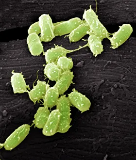
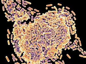
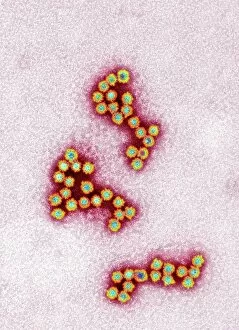
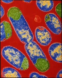
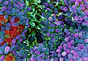
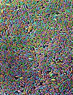
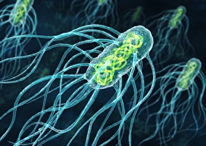
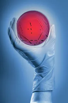
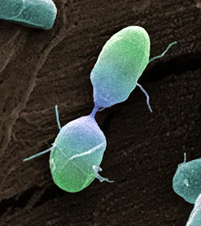
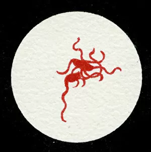
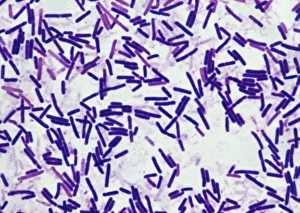
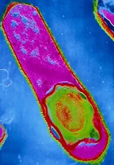
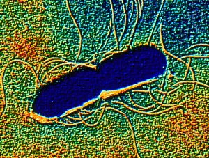
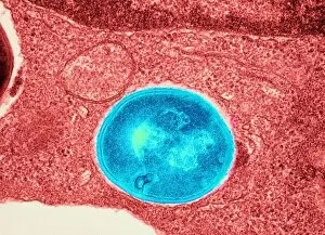
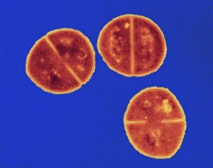
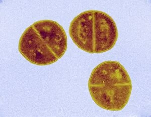
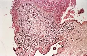
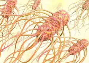
"Unseen Culprits: Exploring the Microscopic World of Food Poisoning" E. Coli bacteria, as seen under a scanning electron microscope (SEM), are one of the leading causes worldwide. Salmonella bacteria, also observed through SEM, pose a significant threat to food safety and can cause severe illness if ingested. The transmission of Norovirus particles, captured using a transmission electron microscope (TEM), is another common source of foodborne illnesses. E. Coli bacteria are notorious for contaminating various foods such as raw meats and unpasteurized dairy products, making proper cooking and hygiene practices crucial in preventing infections. Another SEM image reveals the presence of Salmonella bacteria on contaminated surfaces or in improperly handled food items. A culture displaying Salmonella bacterium highlights the need for strict sanitation measures during food processing to prevent bacterial growth and subsequent contamination. The strain E. coli 0157:H7 is particularly dangerous due to its ability to produce toxins that can lead to severe complications like kidney failure when consumed through contaminated foods or water sources. Witnessed under SEM once again, a dividing Salmonella bacterium showcases their rapid multiplication potential within our digestive system if we consume contaminated foods or beverages. SEM images further reveal the intricate structure of Salmonella typhimurium bacteria responsible for causing gastroenteritis symptoms such as diarrhea, abdominal pain, and fever upon ingestion. Salmonella typhimurium bacterium magnified by an SEM reminds us that even microscopic organisms have substantial impacts on public health; thus ensuring proper food handling practices remains essential in safeguarding against these harmful pathogens.