
Freeze Fracture Collection
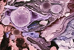
"Unlocking the Intricacies of Freeze Fracture: Revealing the Hidden World in Exquisite Detail" Freeze fracture, a remarkable technique employed in electron microscopy


All Professionally Made to Order for Quick Shipping
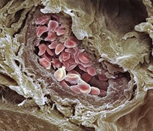
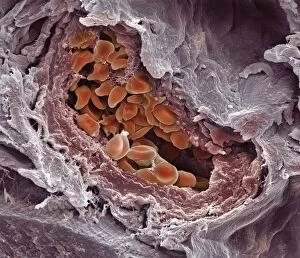
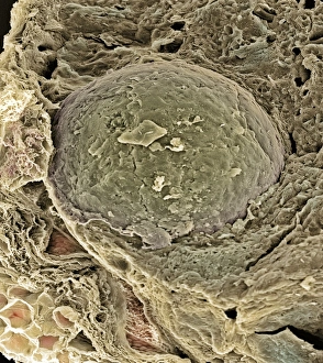
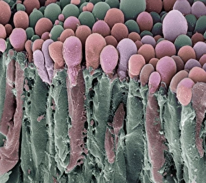
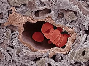
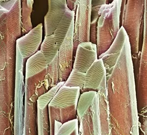
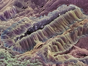
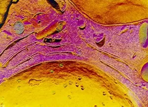
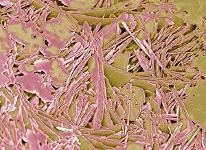
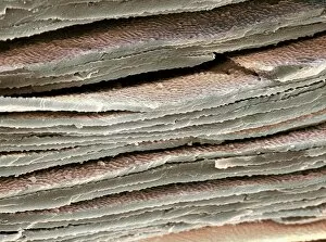
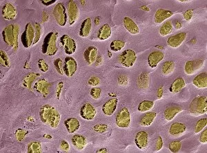
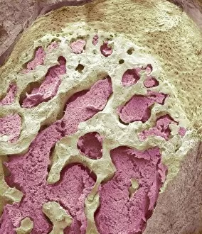
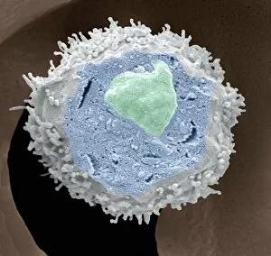
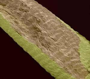
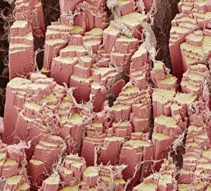
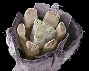
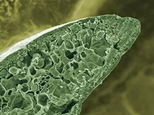
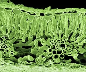
"Unlocking the Intricacies of Freeze Fracture: Revealing the Hidden World in Exquisite Detail" Freeze fracture, a remarkable technique employed in electron microscopy, allows us to delve into the mesmerizing intricacies of various biological structures. From skin layers to chloroplasts, this method offers an unprecedented glimpse into their hidden realms. Peering through the lens of a scanning electron microscope (SEM), we witness the astonishingly complex architecture of skin layers. Each freeze-fractured section unveils a mosaic-like pattern, showcasing the delicate arrangement and organization that lies beneath our outermost protective shield. Moving on to chloroplasts – nature's solar powerhouses – SEM reveals their intricate membrane systems with unparalleled precision. The freeze-fractured images expose these green organelles as a network of interconnected sacs and tubules, highlighting their crucial role in photosynthesis. Intriguingly, even everyday products like baby cream can be subjected to freeze fracture analysis. SEM captures stunning micrographs that showcase its unique composition at an incredibly detailed level. With images such as C017 / 7133, C017 / 7134, and C017 / 7135 revealing frozen snapshots of this cosmetic marvel from different angles, we gain insights into its structural properties. Delving deeper into our circulatory system, blood-filled arteries come under scrutiny through freeze fracture imaging using SEM. Astonishingly detailed visuals like C013 / 7117 and C013 / 7118 allow us to explore these vital conduits for oxygenated blood with awe-inspiring clarity. Every twist and turn within these vessels is laid bare before our eyes. The exploration doesn't stop there; hair follicles become subjects of fascination when observed through SEM after undergoing freeze fracture techniques. These microscopic wonders reveal themselves as intricate tunnels housing hair growth machinery deep within our scalps. Zooming further down to cellular levels brings us face-to-face with egg cells.