Fraud Blocker Skip to main content

Freeze Fractured Collection (page 2)

"Exploring the Intricate World of Freeze-Fractured Baby Cream and Skin Structures" In the realm of scientific imaging

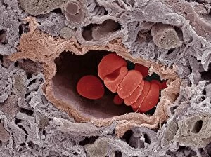
Background imageFreeze Fractured Collection: Coronary capillary, SEM

Coronary capillary, SEM
Coronary capillary. Coloured scanning electron micrograph (SEM) of a section through a freeze- fractured coronary capillary blood vessel

Background imageFreeze Fractured Collection: Ovarian artery, SEM

Ovarian artery, SEM
Ovarian artery. Coloured scanning electron micrograph (SEM) of an ovarian artery that has been freeze-fractured to show internal details

Background imageFreeze Fractured Collection: Arteriole and red blood cells, SEM

Arteriole and red blood cells, SEM
Arteriole. Coloured scanning electron micrograph (SEM) of a cross-section through a small, thin- walled artery known as an arteriole

Background imageFreeze Fractured Collection: Brain artery, SEM

Brain artery, SEM
Brain artery. Coloured scanning electron micrograph (SEM) of a freeze-fractured artery from the brain. The fracture plane has passed through the artery

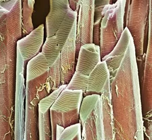
Background imageFreeze Fractured Collection: Muscle fibres, SEM

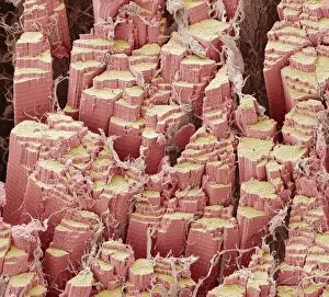
Muscle fibres, SEM
Skeletal muscle fibres. Coloured scanning electron micrograph (SEM) of a freeze-fractured bundle of skeletal (or striated) muscle fibres

Background imageFreeze Fractured Collection: Ulcerative colitis, SEM

Ulcerative colitis, SEM
Ulcerative colitis. Coloured scanning electron micrograph (SEM) of freeze-fractured bowel tissue from a patient suffering from ulcerative colitis

Background imageFreeze Fractured Collection: Eye lens cells, SEM

Eye lens cells, SEM
Eye lens. Coloured scanning electron micrograph (SEM) of a freeze-fractured lens (crystalline lens) of an eye, showing stacked cuboidal epithelium cells

Background imageFreeze Fractured Collection: Hyaline cartilage, SEM

Hyaline cartilage, SEM
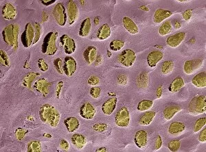
Hyaline cartilage. Coloured scanning electron micrograph (SEM) of a freeze-fractured section through hyaline cartilage, a semi-rigid connective tissue

Background imageFreeze Fractured Collection: Nasturtium stem, SEM

Nasturtium stem, SEM
Nasturtium stem. Coloured scanning electron micrograph (SEM) of a freeze-fractured Nasturtium (Tropaeolum sp.) stem, showing numerous vascular bundles (such as at upper centre)

Background imageFreeze Fractured Collection: Primate finger bone, SEM

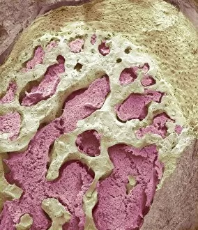
Primate finger bone, SEM

Background imageFreeze Fractured Collection: Primate finger muscle, SEM

Primate finger muscle, SEM

Background imageFreeze Fractured Collection: Lymphocyte white blood cell, SEM

Lymphocyte white blood cell, SEM
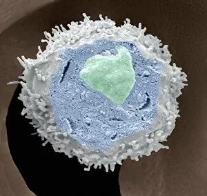
Lymphocyte white blood cell. Coloured scanning electron micrograph (SEM) of a section through a lymphocyte, showing the cell nucleus (upper centre) and cytosol (blue)

Background imageFreeze Fractured Collection: Primate ear canal, SEM

Primate ear canal, SEM

Background imageFreeze Fractured Collection: Primate hand tendon, SEM

Primate hand tendon, SEM

Background imageFreeze Fractured Collection: Nerve bundle, SEM

Nerve bundle, SEM
Nerve bundle. Coloured scanning electron micrograph (SEM) of a freeze-fractured section through a bundle of myelinated nerve fibres. Myelin sheaths (yellow) can be seen surrounding the axons (blue)

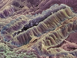
Background imageFreeze Fractured Collection: Muscle fibre, SEM

Muscle fibre, SEM
Muscle fibre. Coloured scanning electron micrograph (SEM) of a freeze-fractured skeletal (or striated) muscle fibre. The fracturing of the fibre has revealed that it consists of a bundle of smaller

Background imageFreeze Fractured Collection: Bird red blood cells, SEM

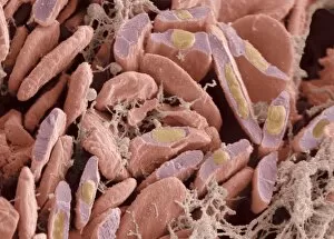
Bird red blood cells, SEM
Bird red blood cells. Coloured scanning electron micrograph (SEM) of fractured red blood cells (erythrocytes, red) from avian heart tissue

Background imageFreeze Fractured Collection: Nettle leaf trichomes, SEM

Nettle leaf trichomes, SEM
Stinging nettle (Urtica dioica) leaf trichomes, coloured scanning electron micrograph (SEM). Trichomes (hair-like) are seen on the upper leaf surface

Background imageFreeze Fractured Collection: Monkey puzzle leaf, SEM

Monkey puzzle leaf, SEM
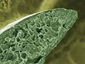
Monkey puzzle leaf. Coloured scanning electron micrograph (SEM) of a freeze-fracture of a leaf of a monkey puzzle tree (Araucaria araucana)

Background imageFreeze Fractured Collection: Birch leaf, SEM

Birch leaf, SEM
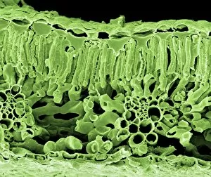
Birch leaf. Coloured scanning electron micrograph (SEM) of a freeze-fracture of a leaf from a birch tree (Betula sp.). The fracture has passed through the leaf

Background imageFreeze Fractured Collection: Lily leaf, SEM

Lily leaf, SEM
Lily leaf. Coloured scanning electron micrograph (SEM) of a freeze-fracture of a leaf from a white water lily (Nymphaea alba)

Background imageFreeze Fractured Collection: Intestinal blood vessels, SEM

Intestinal blood vessels, SEM
Intestinal blood vessels. Coloured scanning electron micrograph (SEM) of a freeze fracture section through the lining of the colon

Background imageFreeze Fractured Collection: Grass leaf, SEM

Grass leaf, SEM
Grass leaf. Coloured scanning electron micrograph (SEM) of a freeze-fracture through a grass leaf. Numerous vascular bundles are present (pale blue)

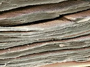
Background imageFreeze Fractured Collection: Tendon fibres, SEM

Tendon fibres, SEM
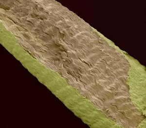
Tendon fibres. Coloured freeze-fracture scanning electron micrograph (SEM) of tendon fibres. These fibres are made from collagen

Background imageFreeze Fractured Collection: Reptilian red blood cells, SEM

Reptilian red blood cells, SEM
Reptilian red blood cells. Coloured scanning electron micrograph (SEM) of whole and fractured red blood cells (erythrocytes, purple) from reptilian tissue

Background imageFreeze Fractured Collection: Gecko retina, SEM

Gecko retina, SEM
Gecko retina. Coloured scanning electron micrograph (SEM) of an oblique freeze-fracture through the retina of a gecko (family Gekkonidae)

Background imageFreeze Fractured Collection: Eye lens fibres, SEM

Eye lens fibres, SEM
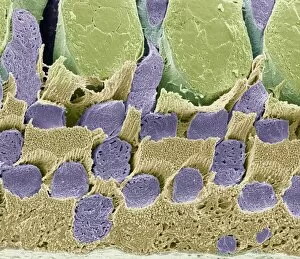
Eye lens fibres. Coloured scanning electron micrograph (SEM) of a freeze-fracture of a lens of an eye, showing the regular arrangement of fibres

Background imageFreeze Fractured Collection: Sectioned leaf, SEM

Sectioned leaf, SEM
Leaf tissue structure. Coloured scanning electron micrograph (SEM) of a freeze-fracture through a rose plant (Rosa sp. ) leaf. The leaf cells are the rectangular shapes in the fracture plane

Background imageFreeze Fractured Collection: Sage leaf oil glands, SEM

Sage leaf oil glands, SEM
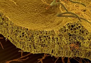
Sage leaf oil glands. Coloured scanning electron micrograph (SEM) of a freeze-fracture through a sage leaf (Salvia officinalis), showing hairs, or trichomes, (green, thin) and oil glands (red)

Background imageFreeze Fractured Collection: Leaf structure, SEM

Leaf structure, SEM
Leaf structure. Coloured scanning electron micrograph (SEM) of a freeze-fracture through a seedling leaf. The top of the leaf is at bottom

Background imageFreeze Fractured Collection: Coiled leaf, SEM

Coiled leaf, SEM
Coiled leaf. Coloured scanning electron micrograph (SEM) of a freeze-fracture through a coiled leaf from a water lily plant (family Nymphaeceae)

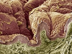
Background imageFreeze Fractured Collection: Skin tissue, SEM

Skin tissue, SEM
Skin tissue. Coloured scanning electron micrograph (SEM) of a freeze-fracture through human skin tissue. The fracture plane (lower frame) has revealed the pseudo-stratified epithelium (below surface)

Background imageFreeze Fractured Collection: Rosemary leaf structure, SEM

Rosemary leaf structure, SEM
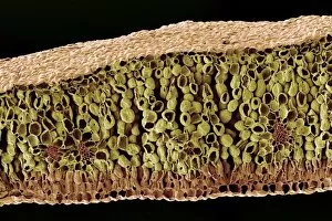
Rosemary leaf. Coloured scanning electron micrograph (SEM) of a freeze-fracture through a leaf from a rosemary plant (Rosmarinus officinalis). The fracture has revealed vascular tissue (green)

Background imageFreeze Fractured Collection: Pancreas acinus, SEM

Pancreas acinus, SEM
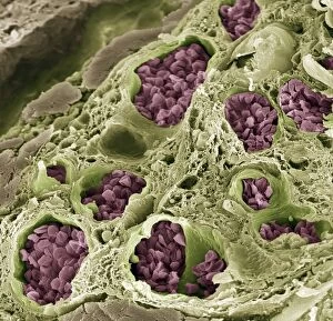
Pancreas acinus. Coloured scanning electron micrograph (SEM) of a freeze-fracture through an acinus (yellow) in the pancreas. An acinus is a collection of glandular epithelial cells

Background imageFreeze Fractured Collection: Artificial sweetener crystals, SEM

Artificial sweetener crystals, SEM
Artificial sweetener crystals. Coloured scanning electron micrograph (SEM) of a freeze-fracture through an artificial sweetener tablet, showing its crystalline structure

Background imageFreeze Fractured Collection: Sugar cube crystals, SEM

Sugar cube crystals, SEM
Sugar cube crystals. Coloured scanning electron micrograph (SEM) of a freeze-fracture through a sugar cube. The fracture plane shows the sucrose crystals that make up the sugar cube


All Professionally Made to Order for Quick Shipping

"Exploring the Intricate World of Freeze-Fractured Baby Cream and Skin Structures" In the realm of scientific imaging, freeze-fracturing offers a fascinating glimpse into the hidden intricacies of various substances and biological structures. Captured under scanning electron microscopy (SEM), these captivating images reveal remarkable details that are otherwise invisible to the naked eye. The first set of images, labeled as "Freeze-fractured baby cream, SEM C017 / 7133, " "Freeze-fractured baby cream, SEM C017 / 7134, " and "Freeze-fractured baby cream, SEM C017 / 7135, " take us on an enchanting journey through this unique product's composition. The frozen fractures expose its internal structure with mesmerizing patterns resembling delicate ice crystals or intricate lacework. These close-ups offer a rare opportunity to appreciate the complexity behind everyday items we often take for granted. Moving beyond skincare products, our exploration continues with stunning visuals captured under SEM. The image titled "Hair follicle, SEM" invites us into the microscopic world beneath our scalps. It unveils hair follicles in astonishing detail – their elongated shapes and distinct layers forming a tapestry that contributes to our individuality. Similarly, we encounter another series labeled as "Skin layers, SEM. " These captivating snapshots showcase different depths within our skin – from epidermis to dermis – each layer playing a crucial role in protecting and nurturing our bodies. Delicate ridges intertwine like miniature mountain ranges while cells appear like tiny building blocks constructing this incredible fortress called skin. Returning once again to hair-related wonders, we delve deeper into hair follicles through multiple images titled "Hair follicle, SEM. " Each photograph unravels new mysteries surrounding these vital structures responsible for hair growth. Their intricate arrangements resemble architectural marvels designed by nature itself.