Fraud Blocker Skip to main content

Fungal Collection (page 8)

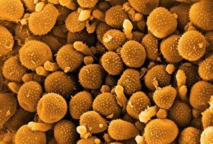
"Fungal Kingdom: Exploring the Intricate World of Fungi" Budding yeast cell: Witness the remarkable process of reproduction as a budding yeast cell emerges

Background imageFungal Collection: Penicillium fungus growing on agar

Penicillium fungus growing on agar
Close-up of a colony of Penicillium chrysogenum fungus growing on agar in a petri dish. This fungus produces the antibiotic penicillin G, seen as small yellow droplets on the surface

Background imageFungal Collection: Neutrophil engulfing fungus, SEM

Neutrophil engulfing fungus, SEM
Neutrophil engulfing fungus. Coloured scanning electron micrograph (SEM) of a neutrophil white blood cell (orange) engulfing and destroying a Candida albicans (thrush) fungal hypha (green)

Background imageFungal Collection: Fungal foot infection, conceptual artwork

Fungal foot infection, conceptual artwork. Single-celled fungi in shape of a footprint. Many fungi live on our body without causing any problems

Background imageFungal Collection: Laetiporus sulphureus

Laetiporus sulphureus
Chicken of the Woods (Laetiporus sulphureus) growing on an oak (Quercus) tree

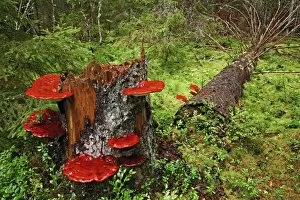
Background imageFungal Collection: Hoof fungus (Fomitopsis pinicola)

Hoof fungus (Fomitopsis pinicola)
A hoof fungus (Fomitopsis pinicola) growing on a tree stump in Georgia, USA

Background imageFungal Collection: Fried quorn

Fried quorn with onions

Background imageFungal Collection: Gill fungus (Lyophyllum decastes)

Gill fungus (Lyophyllum decastes) occurs in dense clusters. The word decastes means a company of ten men. The gills are covered in basidia bearing spores

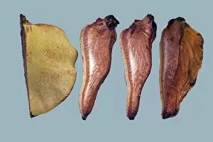
Background imageFungal Collection: Beefsteak Fungus (Fistulina hepatica)

Beefsteak Fungus (Fistulina hepatica). This bracket fungus belonging to the homobasidiomycetes grows on the bark of living oak and sweet chestnut trees

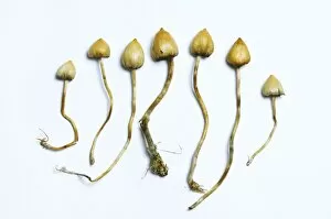
Background imageFungal Collection: Magic mushrooms (Psilocybe semilanceata)

Magic mushrooms (Psilocybe semilanceata). These fungi, commonly known as liberty caps, contain the psychoactive compound psilocybin, which produces a hallucinogenic effect when ingested

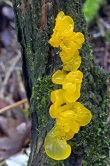
Background imageFungal Collection: Tremella mesenterica

Tremella mesenterica
Yellow brain fungus (Tremella mesenterica) growing on a dead gorse bush. The fruiting body is soft, gelatinous and translucent with brain like lobes

Background imageFungal Collection: Common Earthball (Scleroderma citrinum)

Common Earthball (Scleroderma citrinum). The fruiting body is hemispherical with a tough outer layer covered in scales. Inside the ball is a purple-brown mass (gleba)

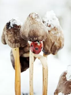
Background imageFungal Collection: Ladybird on fungus

Ladybird on fungus. Seven-spot ladybird (Coccinella septem-punctata) climbing up the stalk of a fungal fruiting body. This is a wood-rotting fungus growing on a dead tree stump

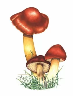
Background imageFungal Collection: Crimson waxcap mushrooms, artwork

Crimson waxcap mushrooms, artwork
Crimson waxcap (Hygrocybe punicea) mushrooms, artwork. This fungus is found in northern Europe. The mushrooms (fruiting bodies) have a bell-shaped cap (lower right)
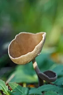
Background imageFungal Collection: Many-coloured polypore fungi

Many-coloured polypore fungi
Many-zoned polypore (Coriolus versicolor) bracket fungi. Wiltshire, UK November 2010

Background imageFungal Collection: Folded rock formation

Folded rock formation. Close-up of folds in a fallen block of rock. These folds are formed as the rock is deformed by immense pressures generated from within the Earths crust as it moves over

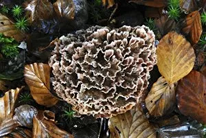
Background imageFungal Collection: Podoscypha fungus

Podoscypha fungus, New Forest, UK October 2009

Background imageFungal Collection: Moulds, historical diagram

Moulds, historical diagram
Zoophytes. Historical diagram drawn to depict moulds as seen by Robert Hooke (1635Ôé¼" 1703) under an early microscope in 1665. Image from La Conquete Du Monde Invisible by Giuseppe Penso

Background imageFungal Collection: ATP sulfurylase molecule

ATP sulfurylase molecule. Computer model showing the structure of an ATP sulfurylase (ATPS) enzyme. ATPS catalyses the primary step of sulphate activation within cells

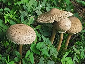
Background imageFungal Collection: Parasol mushroom

Parasol mushroom (Macrolepiota procera). Photographed on Powerstock Common, Dorset, UK, in October

Background imageFungal Collection: Fungal skin infection, artwork

Fungal skin infection, artwork
Fungal skin infection. Artwork of clumps of fungal hyphae (brown), representing an infection of the skin by a fungus. In humans, this is invariably a dermatophyte

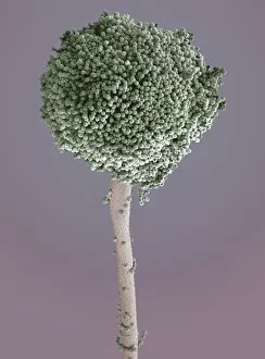
Background imageFungal Collection: Aspergillus fungus, SEM

Aspergillus fungus, SEM
Aspergillus fungus. Coloured scanning electron micrograph (SEM) of the fruiting body (round, top) of a typical mould (Aspergillus sp.)

Background imageFungal Collection: Woman holding split puffball fungus

Woman holding split puffball fungus
MODEL RELEASED. Woman holding a split puffball fungus. The outer wall of this fruiting body has split open to allow the dispersal of the reproductive spores from the sac inside

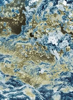
Background imageFungal Collection: Stilton cheese, SEM

Stilton cheese, SEM
Stilton. Coloured scanning electron micrograph (SEM) of a blue vein in a piece of Stilton cheese. The colour and flavour of the cheese is produced by the fungus Penicillium roquefortii

Background imageFungal Collection: Curvularia geniculata fungus, SEM

Curvularia geniculata fungus, SEM
Curvularia geniculata fungus. Coloured scanning electron micrograph (SEM) of the fruiting body of the mould fungus Curvularia geniculata

Background imageFungal Collection: Nematode-killing fungus, SEM

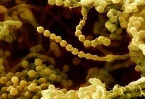
Nematode-killing fungus, SEM
Nematode-killing fungus. Coloured scanning electron micrograph (SEM) of spore chains from the fungus Paecilomyces variotii. This fungus is found in soil and decaying plant matter

Background imageFungal Collection: Dead mans fingers (Xylaria polymorpha)

Dead mans fingers (Xylaria polymorpha) fungi. This inedible fungus occurs on decaying wood. The fruiting body is hard and varies in shape from cylindrical to knobbly

Background imageFungal Collection: Bracket fungus (Laetiporus sulphureus)

Bracket fungus (Laetiporus sulphureus)
Bracket fungus. Young sulphur-coloured fruiting body of the edible bracket fungus known as the chicken of the woods (Laetiporus sulphureus), growing on a living willow tree

Background imageFungal Collection: Bracket fungus (Daedaleopsis confragosa)

Bracket fungus (Daedaleopsis confragosa)
Blushing bracket fungus (Daedaleopsis confragosa). Close-up of the underside of the blushing bracket fungus showing its pores. The pores are the ends of the tubes within which the spores are produced

Background imageFungal Collection: Bracket fungus (Polyporus squamosus)

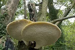
Bracket fungus (Polyporus squamosus)
Bracket fungus. Dryads saddle (Polyporus squamosus) bracket fungus on a tree

Background imageFungal Collection: Parasol mushrooms

Parasol mushrooms (Macrolepiota procera). Photographed in Narke, Sweden

Background imageFungal Collection: Psathyrella mushrooms after forest fire

Psathyrella mushrooms after forest fire
Psathyrella pennata mushrooms growing one year after a forest fire. This fire took place in a clear-felled area in the Tiveden forest, in Vastra Gotaland County, Sweden

Background imageFungal Collection: Geopyxis elf-cup fungi after forest fire

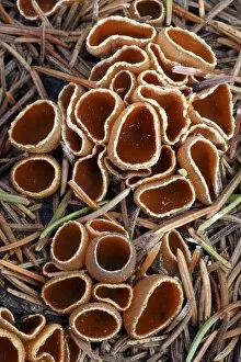
Geopyxis elf-cup fungi after forest fire
Geopyxis carbonaria elf-cup fungi growing one year after a forest fire. This fire took place in a clear-felled area in the Tiveden forest, in Vastra Gotaland County, Sweden

Background imageFungal Collection: Reishi fungus

Reishi fungus
Reishe fungus (Ganoderma lucidum) growing on a dead tree. Photographed in Narke, Sweden

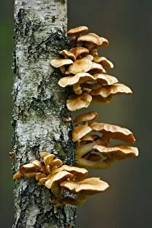
Background imageFungal Collection: Honey fungus

Honey fungus (Armillaria mellea) growing on a tree trunk. Photogroahed in Narke, Sweden

Background imageFungal Collection: Jersey cow bolete

Jersey cow bolete (Suillus bovinus). Photographed in Narke, Sweden

Background imageFungal Collection: Myxomphalia mushroom after forest fire

Myxomphalia mushroom after forest fire
Myxomphalia maura mushrooms growing one year after a forest fire. This fire took place in a clear-felled area in the Tiveden forest, in Vastra Gotaland County, Sweden

Background imageFungal Collection: Helvella macropus mushroom

Helvella macropus mushroom
Helvella macropus fungus fruiting bodies. Photographed in Narke, Sweden

Background imageFungal Collection: Hares ear fungus

Hares ear fungus (Otidea onotica). Photographed in Narke, Sweden

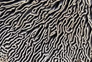
Background imageFungal Collection: Maze-gill fungus underside

Maze-gill fungus underside
Maze-gill fungus (Daedalea quercina) underside, showing the gills where the reproductive spores (not seen) are released. Photographed in October, on Powerstock Common, Dorset, UK

Background imageFungal Collection: Peziza cup fungus after forest fire

Peziza cup fungus after forest fire
Peziza cup fungus (Peziza lobulata) growing two months after a forest fire. This fire took place in a clear-felled area in the Tiveden forest, in Vastra Gotaland County, Sweden

Background imageFungal Collection: Plant regrowth after forest fire

Plant regrowth after forest fire
Plant regrowth one year after a forest fire. The plants and fungi seen here include Pholiota highlandensis mushrooms (lower left), the common liverwort (Marchantia polymorpha, white-green, centre)

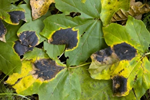
Background imageFungal Collection: Tar Spot Fungus (Rhytisma acerinum)

Tar Spot Fungus (Rhytisma acerinum) on the leaves of a Field Maple (Acer campestre) tree. Photographed in autumn in Romania

Background imageFungal Collection: Birch bolete (Leccinum scabrum) mushroom

Birch bolete (Leccinum scabrum) mushroom
Brown birch bolete (Leccinum scabrum) mushroom growing amongst common heather (Calluna vulgaris). Photographed in the Cairngorms National Park, Scotland, in August

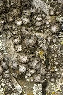
Background imageFungal Collection: Lichen (Sticta sylvatica)

Lichen (Sticta sylvatica) growing on rocks. Photographed in Auvergne in France

Background imageFungal Collection: Bean rust pustules, SEM

Bean rust pustules, SEM
Bean rust (Uromyces sp.) pustules, coloured scanning electron micrograph (SEM). The spores (orange) are emerging from pustules on the underside of a leaf on a bean plant

Background imageFungal Collection: Mushroom gill cap, light micrograph

Mushroom gill cap, light micrograph. Transverse section through the cap of a common mushroom (Psalliota sp.) showing the gill lamellae (white)

Background imageFungal Collection: Bean rust spores, SEM

Bean rust spores, SEM
Bean rust (Uromyces sp.) spores, coloured scanning electron micrograph (SEM). Bean rust is a fungal disease that infects bean plants

Background imageFungal Collection: Rust fungus infection, light micrograph

Rust fungus infection, light micrograph. Transverse section through a barberry (Berberis sp.) leaf infected by a parasitic rust (Puccinia sp.)


All Professionally Made to Order for Quick Shipping

"Fungal Kingdom: Exploring the Intricate World of Fungi" Budding yeast cell: Witness the remarkable process of reproduction as a budding yeast cell emerges, ready to create new life. Fly agaric mushrooms: Delve into the enchanting realm of fly agaric mushrooms, with their vibrant red caps and white spots, captivating both nature enthusiasts and fairytale lovers alike. SEM of penicillin fungus: Uncover the microscopic beauty of penicillin fungus through scanning electron microscopy (SEM), revealing its intricate structures that have revolutionized medicine. Culture of Aspergillus nidulans fungus: Step into a laboratory where cultures of Aspergillus nidulans fungus thrive, showcasing their unique growth patterns and potential for scientific research. Cep mushroom (Boletus edulis): Discover the culinary delight known as cep mushroom or Boletus edulis, renowned for its rich flavor and sought-after by chefs around the world. Candida fungus, SEM: Explore the unseen world within our bodies as Candida fungi are magnified under an electron microscope, shedding light on their role in various infections and diseases. Mushroom gills, SEM: Marvel at the intricate network of gills found beneath mushroom caps when observed through scanning electron microscopy (SEM), highlighting their crucial role in spore production. Fly agaric fungi: Journey through enchanted forests where fly agaric fungi flourish; these iconic red-and-white toadstools evoke a sense of wonderment while harboring fascinating ecological relationships with other organisms. Dividing yeast cells, SEM: Peer into a microscopic universe where dividing yeast cells undergo mitosis under high-resolution imaging techniques—witnessing life's continuous cycle unfold before your eyes. Penicillium roqueforti fungus: Meet Penicillium roqueforti—a blue mold used in cheese-making—whose distinctive flavor and appearance contribute to the creation of beloved blue cheeses worldwide.