
Ganglion Collection
Ganglion: The Nexus of Chakras and the Nervous System In the intricate web of our body's functioning


All Professionally Made to Order for Quick Shipping
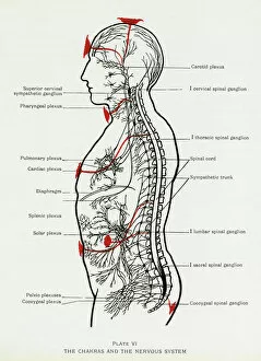
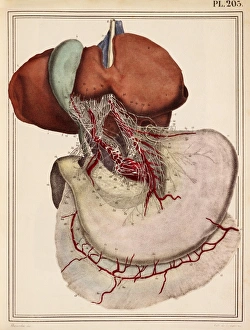
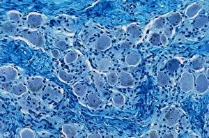
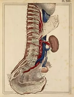
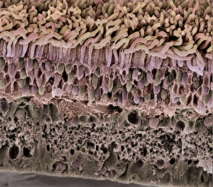
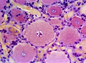
Ganglion: The Nexus of Chakras and the Nervous System In the intricate web of our body's functioning, ganglions play a crucial role in connecting various aspects of our anatomy. These clusters of nerve cell bodies are like energetic hubs, bridging the realms of chakras and the nervous system. A normal side view of an adult skull reveals a fascinating sight—the spinal cord gracefully nestled within its protective bony structure, and is here that ganglia can be found, acting as vital intermediaries between our brain and peripheral nerves. Illustrations showcasing peripheral nerves offer us a glimpse into their complexity. Ganglia stand out prominently, surrounded by nerve fibers intricately woven together like delicate threads, and are accompanied by myelin sheaths, which provide insulation to these neural pathways. Arteries and veins meander alongside them while fat cells cushion this remarkable network. Even specific organs have their own unique ganglia systems. Take for instance the gall bladder—a small but significant organ involved in digestion—its anatomy includes specialized ganglia that contribute to its proper functioning. Sometimes nature surprises us with unexpected encounters, such as when a bee sting remains embedded in human skin. A cross-section model allows us to witness how even this seemingly minor event impacts our nervous system at the site where it occurred. The interconnectedness continues throughout our body's landscape; artwork from 1825 depicts nerves extending towards vital organs like the liver and stomach. These illustrations remind us that every bodily function relies on precise communication facilitated by intricate networks of ganglia. Microscopic views bring forth astonishing details about these neural structures. Light micrographs reveal nerve ganglia bathed in ethereal hues, highlighting their significance in transmitting information across vast distances within our bodies. Zooming further into cellular levels through transmission electron microscopy (TEM), we encounter smooth muscle cells lining gut walls—an integral part regulated by myenteric nerve plexus containing numerous ganglia.