
Gastroenterology Collection
"Gastroenterology: Exploring the Intricacies of the Human Digestive System Through Art" Delve into the captivating world of gastroenterology


All Professionally Made to Order for Quick Shipping
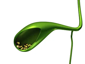
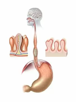
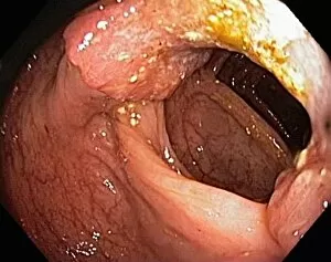
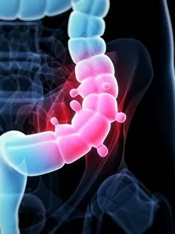
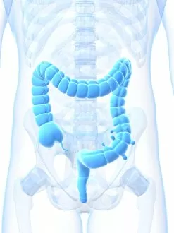
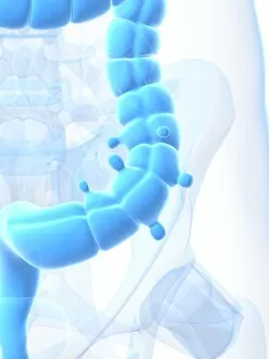
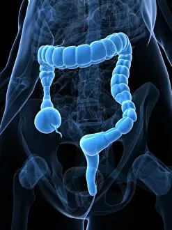
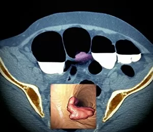
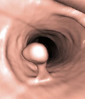
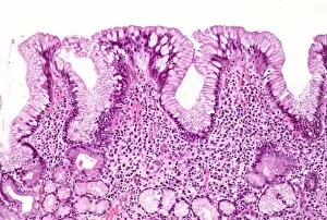
"Gastroenterology: Exploring the Intricacies of the Human Digestive System Through Art" Delve into the captivating world of gastroenterology, where art meets science to unravel the mysteries within our digestive system. From gallstones to colorectal cancer cells, each artwork offers a unique glimpse into these intricate processes. Marvel at the stunning depiction of gallstones, crystalline formations that can cause discomfort and pain if left untreated. Witness how these tiny yet impactful structures disrupt the delicate balance within our bodies. Embark on a visual journey through an artistic representation of the human digestive system. Admire its complexity and marvel at how it seamlessly functions to nourish us every day. Explore Gastric Antral Vascular Ectasia (GAVE), also known as watermelon stomach, depicted in mesmerizing detail. Discover this rare condition characterized by dilated blood vessels in the stomach lining, causing chronic gastrointestinal bleeding. Observe an exquisite rendering of colorectal cancer cells under a scanning electron microscope (SEM). Gain insight into this devastating disease and appreciate scientific advancements aimed at combating its progression. Contemplate Ulcerative Pancolitis, portrayed with striking imagery capturing its effects on intestinal health. Reflect upon this inflammatory bowel disease's impact on individuals' lives and ongoing efforts towards finding effective treatments. Immerse yourself in thought-provoking artwork depicting Toxic Megacolon—a potentially life-threatening condition marked by severe inflammation and dilation of the colon. Contemplate both its destructive nature and medical interventions designed to save lives. Experience conceptual artwork illustrating abdominal pain—an enigmatic sensation often associated with various gastrointestinal disorders. Engage your senses as you ponder over potential causes while appreciating medicine's tireless pursuit for relief. Witness an evocative portrayal showcasing constipated colon—depicting both physical discomfort and emotional strain endured by those affected. Acknowledge that even seemingly mundane conditions can significantly impact one's well-being.