
Gastrointestinal Tract Collection (#3)
The gastrointestinal tract, also known as the digestive system, is a complex network of organs that play a crucial role in our overall health


All Professionally Made to Order for Quick Shipping
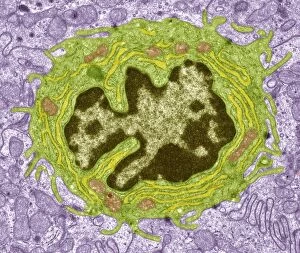
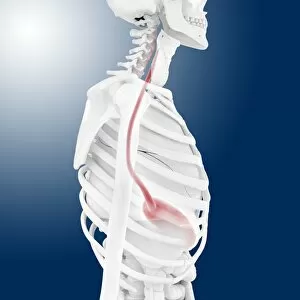
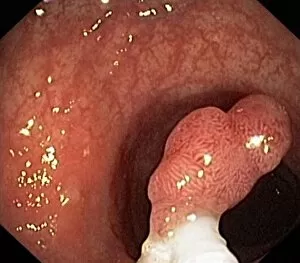
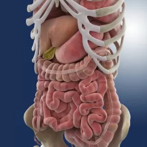
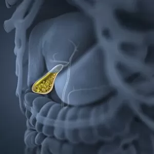
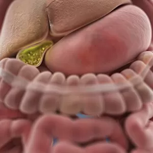
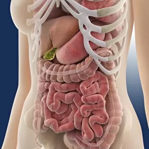
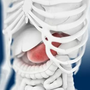
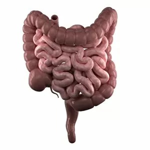
The gastrointestinal tract, also known as the digestive system, is a complex network of organs that play a crucial role in our overall health. From the mouth to the anus, this intricate system ensures that we can properly digest and absorb nutrients from the food we consume. One important organ within this system is the appendix. In a medical illustration depicting appendicitis, we can see how inflammation of this small pouch attached to the large intestine can cause severe pain and discomfort. Another fascinating aspect of the gastrointestinal tract is found in its microscopic structures. Intestinal microvilli, shown through a transmission electron microscope (TEM), are tiny finger-like projections lining the walls of our intestines. These microvilli greatly increase surface area for nutrient absorption. When it comes to understanding how our internal organs function together, visual aids are incredibly helpful. A female body illustration showcases both her digestive and circulatory systems side by side, highlighting their interconnectedness. Similarly, an anatomy diagram reveals not only male respiratory organs but also other internal structures vital for proper bodily functions. This comprehensive view emphasizes how different systems work harmoniously within our bodies. In another 3D rendering featuring healthy female internal organs, we gain insight into their spatial arrangement and proximity to one another. Such visuals aid medical professionals in diagnosing potential issues or diseases affecting these areas. A male anatomy diagram further highlights various internal organs responsible for digestion and respiration processes. Understanding their locations helps us appreciate their roles in maintaining optimal health. Looking at human digestive system anatomy from a front view provides valuable insights into its complexity and organization. With each component labeled accordingly - from esophagus to rectum - it becomes easier to comprehend its intricacies. Superimposing interior organ images onto a woman's midsection offers an even clearer picture of where these vital structures reside within her body. This visualization aids doctors when discussing specific conditions or treatment plans with patients. Lastly, a blue background showcasing a male skeleton with internal organs reminds us of the importance of maintaining a healthy gastrointestinal tract.