
Golgi Body Collection
The Golgi body, also known as the Golgi apparatus, is a vital organelle found in both animal and plant cells


All Professionally Made to Order for Quick Shipping
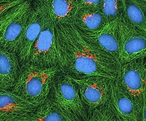
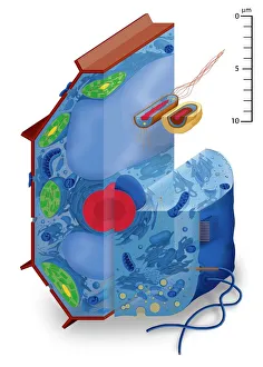
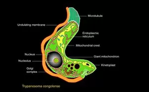
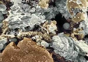
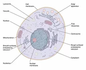
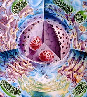
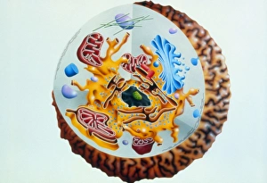
The Golgi body, also known as the Golgi apparatus, is a vital organelle found in both animal and plant cells. It plays a crucial role in processing and packaging proteins before they are transported to their final destinations within the cell or outside of it. In HeLa cells, which were derived from cervical cancer cells taken from Henrietta Lacks, the they are be visualized using a light micrograph labeled C017 / 8298. This image allows scientists to study its structure and function within these unique cell types. Artwork depicting different cell types showcases the presence of the Golgi body in various organisms. For instance, an artwork featuring Trypanosome protozoan highlights how this single-celled organism possesses a distinct Golgi apparatus that aids in protein modification and secretion. A scanning electron micrograph (SEM) provides an intricate view of the Golgi apparatus's ultrastructure within an animal cell. This organelle appears as stacked flattened sacs called cisternae interconnected by tubules, giving it a distinctive appearance under high magnification. When examining animal cell structures using models, one can observe the presence of several organelles alongside the Golgi body. These include the nucleus, lysosomes, centrioles, mitochondria, endoplasmic reticulum (ER), ribosomes, cytoplasm filled with vesicles for transport processes, thin plasma membrane enclosing everything inside and microvilli projecting at its top surface for increased surface area absorption. Similarly depicted through artwork is another model showcasing plant cells' complexity where you can find not only chloroplasts but also a well-defined Golgi apparatus involved in synthesizing complex carbohydrates like cellulose for building sturdy cell walls. Illustrations such as C018 / 0734 provide detailed depictions of animal cells highlighting key features like nuclei and prominently displaying the intricate network formed by numerous stacks of cisternae making up the Golgi apparatus.