Fraud Blocker Skip to main content

Gram Negative Collection

"Exploring the Intricate World of Gram-Negative Bacteria

Background imageGram Negative Collection: E. coli bacteria, SEM

E. coli bacteria, SEM
E. coli bacteria. Coloured scanning electron micrograph (SEM) of Escherichia coli bacteria. These bacteria are a normal part of the intestinal flora in humans and other animals

Background imageGram Negative Collection: Salmonella bacteria, SEM

Salmonella bacteria, SEM
Salmonella bacteria, coloured scanning electron micrograph (SEM). These Gram-negative rod-shaped bacteria can cause food poisoning when eaten in contaminated food

Background imageGram Negative Collection: Cell types, artwork

Cell types, artwork
Cell types. Cutaway artwork with a scale bar (upper right, in micrometres) showing the relative sizes of eukaryotic cells (those containing a nucleus) and prokaryotic cells (those lacking a nucleus)

Background imageGram Negative Collection: Pseudomonas aeruginosa bacteria, SEM

Pseudomonas aeruginosa bacteria, SEM
Pseudomonas aeruginosa bacteria, coloured scanning electron micrograph (SEM). These Gram-negative rod-shaped bacteria are found in soil and water, and as normal flora in the human intestine

Background imageGram Negative Collection: E. coli bacteria, SEM

E. coli bacteria, SEM
E. coli bacteria. Coloured scanning electron micrograph (SEM) of Escherichia coli bacteria (purple) taken from the small intestine of a child. E

Background imageGram Negative Collection: Pseudomonas aeruginosa bacteria, SEM

Pseudomonas aeruginosa bacteria, SEM
Pseudomonas aeruginosa bacteria, coloured scanning electron micrograph (SEM). These Gram-negative rod-shaped bacteria are found in soil and water, and as normal flora in the human intestine

Background imageGram Negative Collection: E. coli bacterium, TEM

E. coli bacterium, TEM
E. coli bacterium, coloured transmission electron micrograph (TEM). E. coli are Gram-negative rod-shaped bacteria that are part of the normal flora of the human gut

Background imageGram Negative Collection: Pseudomonas aeruginosa bacteria, SEM

Pseudomonas aeruginosa bacteria, SEM
Pseudomonas aeruginosa bacteria, coloured scanning electron micrograph (SEM). These Gram-negative rod-shaped bacteria are found in soil and water, and as normal flora in the human intestine

Background imageGram Negative Collection: E. coli bacterium, TEM

E. coli bacterium, TEM
E. coli bacterium. Coloured transmission electron micrograph (TEM) of an Escherichia coli bacterium in the early stages of binary fission, the process by which the bacterium divides

Background imageGram Negative Collection: Pseudomonas aeruginosa bacteria, SEM C017 / 7140

Pseudomonas aeruginosa bacteria, SEM C017 / 7140
Pseudomonas aeruginosa bacteria, coloured scanning electron micrograph (SEM). These Gram-negative rod-shaped bacteria are found in soil, water and as normal flora in the human intestine

Background imageGram Negative Collection: Gram-stained bacteria C016 / 7500

Gram-stained bacteria C016 / 7500
Gram-stained bacteria. Computer artwork showing gram-positive (blue) and gram-negative (red) bacteria. Gram-positive bacteria are those that are stained dark blue or violet by Gram staining

Background imageGram Negative Collection: E. coli bacteria, illustration C018 / 0733

E. coli bacteria, illustration C018 / 0733
Escherichia coli, illustration. E. coli are Gram-negative rod-shaped bacteria that are part of the normal flora of the human gut

Background imageGram Negative Collection: E. coli bacteria, SEM C014 / 0385

E. coli bacteria, SEM C014 / 0385
E. coli bacteria. Coloured scanning electron micrograph (SEM) of Escherichia coli (E. coli) bacteria (rod-shaped) found in a urine sample from a patient with a urinary tract infection (UTI)

Background imageGram Negative Collection: E. coli bacteria, SEM C014 / 0386

E. coli bacteria, SEM C014 / 0386
E. coli bacteria. Coloured scanning electron micrograph (SEM) of Escherichia coli (E. coli) bacteria (rod-shaped) found in a urine sample from a patient with a urinary tract infection (UTI)

Background imageGram Negative Collection: Actin-based motility, SEM C017 / 8335

Actin-based motility, SEM C017 / 8335
Actin-based motility. Coloured scanning electron micrograph (SEM) of Shigella sp. bacteria (pink) infecting cells. Filaments of the protein actin, which is part of the cells cystoskeleton

Background imageGram Negative Collection: Actin-based motility, SEM C017 / 8334

Actin-based motility, SEM C017 / 8334
Actin-based motility. Coloured scanning electron micrograph (SEM) of Shigella sp. bacteria (pink) infecting cells. Filaments of the protein actin, which is part of the cells cystoskeleton

Background imageGram Negative Collection: Actin-based motility, light micrograph C017 / 8333

Actin-based motility, light micrograph C017 / 8333
Actin-based motility. 3D model made from optical sections of Shigella sp. bacteria (pink) infecting cells. Filaments of the protein actin, which is part of the cells cystoskeleton

Background imageGram Negative Collection: Q fever bacteria, SEM C017 / 8303

Q fever bacteria, SEM C017 / 8303
Q fever bacteria. Coloured scanning electron micrograph (SEM) of a fracture through a vacuole in a Vero cell showing replicating Coxiella burnetii bacteria (yellow)

Background imageGram Negative Collection: Acetobacter bacteria, SEM

Acetobacter bacteria, SEM
Vinegar-forming bacteria. Coloured scanning electron micrograph (SEM) of Acetobacter aceti bacteria, which are used in the production of vinegar

Background imageGram Negative Collection: Neutrophils and Shigella bacteria, SEM

Neutrophils and Shigella bacteria, SEM
Neutrophils and Shigella bacteria. Coloured scanning electron micrograph (SEM) of neutrophil white blood cells (large, yellow-green) and Shigella bacteria (orange, upper right)

Background imageGram Negative Collection: Neutrophil and trapped bacteria, TEM

Neutrophil and trapped bacteria, TEM
Neutrophil and trapped bacteria. Coloured transmission electron micrograph (TEM) of Neisseria gonorrhoeae bacteria (blue) trapped by a neutrophil (orange), a type of white blood cell

Background imageGram Negative Collection: Diagram of cell wall & flagellum of Gram- bacteria

Diagram of cell wall & flagellum of Gram- bacteria
Diagrammatic representation of the cell wall, cell membrane, and a flagellum of a Gram-negative bacterium. Gram-negative bacteria have an outer lipopolysaccharide layer (above, yellow)

Background imageGram Negative Collection: Helicobacter pylori bacterium, artwork

Helicobacter pylori bacterium, artwork

Background imageGram Negative Collection: Acinetobacter bacteria, artwork

Acinetobacter bacteria, artwork

Background imageGram Negative Collection: Spirillum bacteria, artwork

Spirillum bacteria, artwork

Background imageGram Negative Collection: E. coli bacterium, computer artwork

E. coli bacterium, computer artwork
E. coli bacterium. Computer artwork of an Escherichia coli bacterium

Background imageGram Negative Collection: Red blood cell and bacteria, SEM

Red blood cell and bacteria, SEM
Red blood cell and bacteria. Coloured scanning electron micrograph (SEM) of a crenated red blood cell (red) surrounded by Escherichia coli (E. coli) bacteria

Background imageGram Negative Collection: Shigella bacteria, SEM

Shigella bacteria, SEM
Shigella bacteria. Scanning electron micrograph (SEM) of Shigella sp. Bacteria (red) on the surface of a cell. Shigella sp

Background imageGram Negative Collection: Gram-negative bacterium, artwork

Gram-negative bacterium, artwork
Gram-negative bacterium. Artwork of a prokaryotic, Gram-negative rod-shaped bacterium with a single polar flagellum (spiral structure, upper left)

Background imageGram Negative Collection: Gram-negative bacteria, artwork

Gram-negative bacteria, artwork
Gram-negative bacteria. Computer artwork showing the cell structure of a gram-negative bacterium. Gram-negative bacteria are bacteria that do not retain crystal violet dye in the Gram staining

Background imageGram Negative Collection: Salmonella bacteria, TEM

Salmonella bacteria, TEM
Salmonella bacteria. Coloured transmission electron micrograph (TEM) of a Salmonella sp. bacteria (green) in the cytoplasm of an infected cell

Background imageGram Negative Collection: Bacterial cell wall comparison, artwork

Bacterial cell wall comparison, artwork
Bacterial cell wall comparison. Computer artwork comparing the structure of the cell wall from a gram-negative bacterium (left) with that of a gram-positive bacterium (right)

Background imageGram Negative Collection: Legionella bacteria, SEM

Legionella bacteria, SEM
Legionella bacteria. Coloured scanning electron micrograph (SEM) of Legionella pneumophila bacteria, the cause of Legionnaires disease

Background imageGram Negative Collection: E. coli bacterium strain O157: H7, TEM

E. coli bacterium strain O157: H7, TEM
E. coli bacterium strain O157:H7, coloured transmission electron micrograph (TEM). E. coli are Gram-negative rod-shaped bacteria that are part of the normal flora of the human gut

Background imageGram Negative Collection: Legionella bacteria, light micrograph

Legionella bacteria, light micrograph
Legionella bacteria. Coloured light micrograph of Legionella pneumophila bacteria, the cause of Legionnaires disease. This rod-shaped

Background imageGram Negative Collection: E. coli bacteria strain O157: H7, TEM

E. coli bacteria strain O157: H7, TEM
E. coli bacteria strain O157:H7, coloured transmission electron micrograph (TEM). E. coli are Gram-negative rod-shaped bacteria that are part of the normal flora of the human gut

Background imageGram Negative Collection: Legionella bacterium, TEM

Legionella bacterium, TEM
Legionella bacteria. Coloured transmission electron micrograph of a Legionella pneumophila bacterium, the cause of Legionnaires disease

Background imageGram Negative Collection: S. maltophilia bacterium, TEM

S. maltophilia bacterium, TEM
Stenotrophomonas maltophilia bacterium, coloured transmission electron micrograph (TEM). This aerobic Gram-negative bacterium, previously known as Pseudomonas maltophilia

Background imageGram Negative Collection: Helicobacter bilis bacteria, SEM

Helicobacter bilis bacteria, SEM
Helicobacter bilis bacteria, coloured field emission scanning electron micrograph (FESEM). H. bilis is a Gram-negative spiral-shaped bacterium found in the intestines of rodents, dogs and cats

Background imageGram Negative Collection: Salmonella bacteria

Salmonella bacteria

Background imageGram Negative Collection: Salmonella bacterium, TEM

Salmonella bacterium, TEM
Salmonella sp. bacterium, coloured transmission electron micrograph (TEM). Salmonella is a Gram-negative rod-shaped bacterium in the same family as Escherichia coli (Enterobacteriaceae)

Background imageGram Negative Collection: Salmonella bacterium

Salmonella bacterium. Coloured transmission electron micrograph (TEM) of a section through a Salmonella sp. bacterium. It is seen on a fractal background


All Professionally Made to Order for Quick Shipping

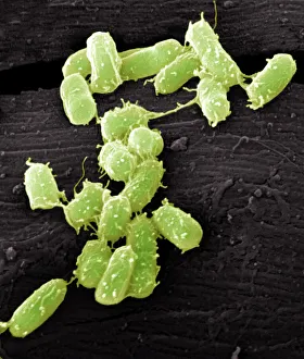
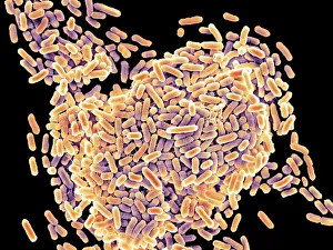
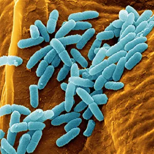
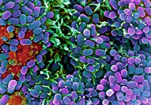
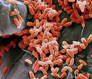
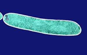
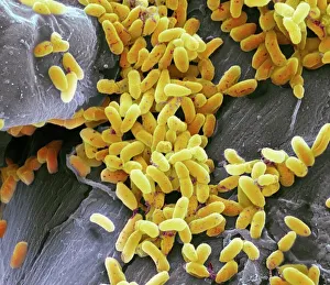
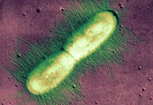
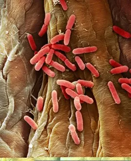
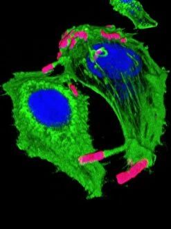
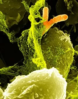
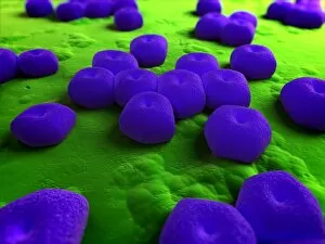
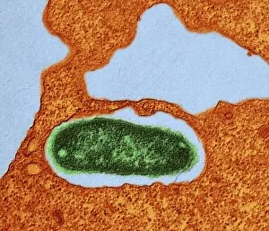
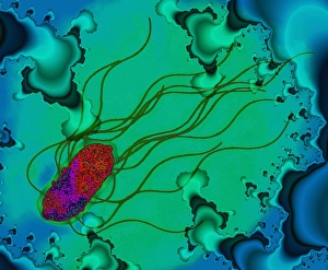
"Exploring the Intricate World of Gram-Negative Bacteria: A Visual Journey" Step into the microscopic realm and witness the fascinating world of gram-negative bacteria through scanning electron microscopy (SEM) and transmission electron microscopy (TEM). First, we encounter E. Coli bacteria, their slender rod-like bodies captured in stunning detail by SEM. These notorious inhabitants of our intestines remind us of their presence and importance in both human health and scientific research. Moving on, Salmonella bacteria come into view under SEM, showcasing their unique shape and surface features. These pathogens are responsible for causing foodborne illnesses but also serve as valuable models for studying bacterial infection mechanisms. But it's not just about disease-causing organisms; artistry can be found even at the cellular level. Admire the intricate artwork created by different cell types - a testament to nature's creativity. Next up is Pseudomonas aeruginosa bacteria seen through SEM, revealing their distinctive appearance with flagella protruding from their surfaces. Known for causing infections in immunocompromised individuals, these versatile microbes have become a focus of intense scientific investigation. Switching to TEM imagery, we zoom in closer on an individual E. Coli bacterium – its internal structures visible with astonishing clarity. This technique allows scientists to delve deeper into understanding cellular processes within these tiny entities. Returning to SEM visuals, Pseudomonas aeruginosa once again takes center stage – this time displaying intricate details that make them stand out among other gram-negative species. TEM provides another glimpse inside an E. coli bacterium - unveiling its inner workings like never before. This high-resolution imaging helps researchers uncover new insights into bacterial physiology and pathogenesis. Lastly, Salmonella typhimurium bacteria appear under SEM magnification – reminding us once more of the complex interplay between humans and microorganisms that can lead to illness or symbiotic relationships. In this captivating visual journey through various techniques, we witness the beauty and complexity of gram-negative bacteria.