
Gynaecology Collection
Gynaecology, the field of medicine that focuses on women's reproductive health, encompasses a wide range of topics and images


All Professionally Made to Order for Quick Shipping
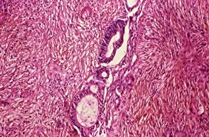
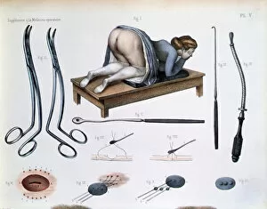
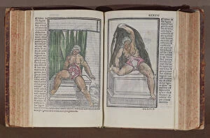
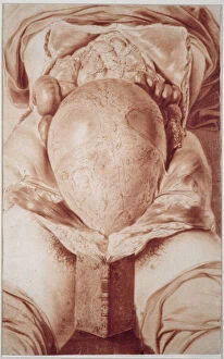
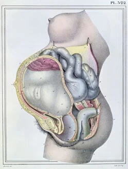
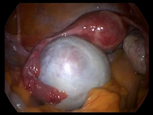
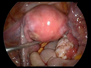
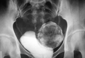
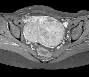
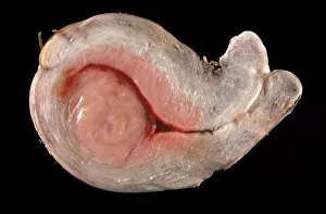
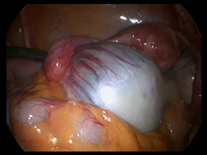
Gynaecology, the field of medicine that focuses on women's reproductive health, encompasses a wide range of topics and images. From the study of ovarian cancer to the intricate details captured in light micrograph C015/7103, gynaecology delves into understanding and treating various conditions affecting women. In historical depictions, we see the significance placed on childbirth and its role in family dynamics. The midwife proudly presenting the first-born male offspring to other family members symbolizes both joy and anticipation for future generations. Ms Hunter's detailed drawings from Anatomy of the Human Gravid Uterus provide valuable insights into pregnancy and fetal development during William Hunter's time. The representation of female genitals on painted cardboard from around 1830 showcases how anatomical knowledge was shared through visual aids. Woodcut prints depicting childbirth highlight both the beauty and challenges associated with bringing new life into this world. Gynaecological procedures are also an essential part of this medical specialty. An operation on a vesicovaginal fistula, as depicted in Jean-Baptiste Marc Bourgery's coloured engraving from Traite Complet de l'Anatomie de l'Homme, demonstrates surgical interventions aimed at improving women's quality of life. Tools like Joseph-Claude-Anthelme Recamier's vaginal speculum further illustrate advancements made in examining female anatomy throughout history. Commentaria cum amplissimis additionibus super anatomiam provides a comprehensive view of female anatomy beyond just reproductive organs. Lastly, a diagram showing stages of embryonic development reminds us that gynaecology encompasses not only adult women but also their potential for motherhood.