
Hematology Collection
Hematology, the study of blood and its components, is a fascinating field that delves into the intricate workings of our circulatory system


All Professionally Made to Order for Quick Shipping
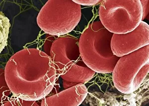
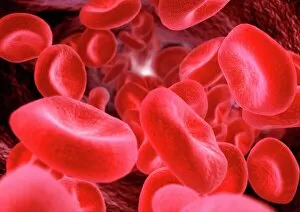
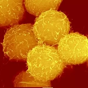
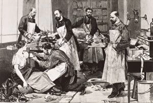
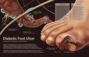
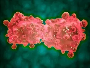
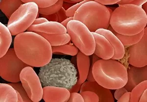
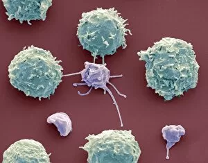
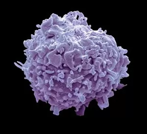
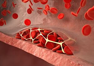
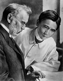
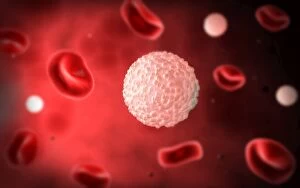
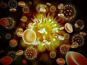
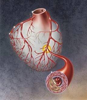
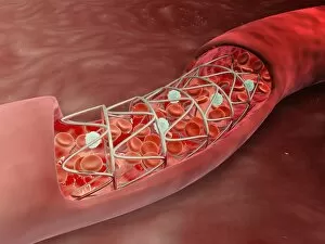
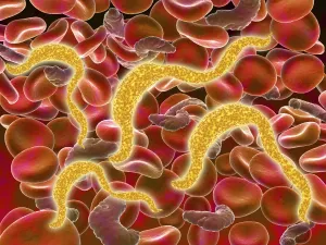
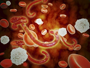
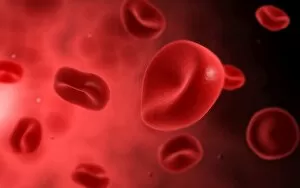
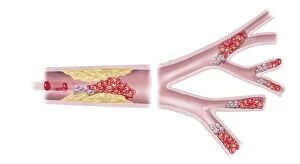
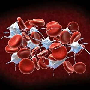
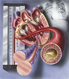
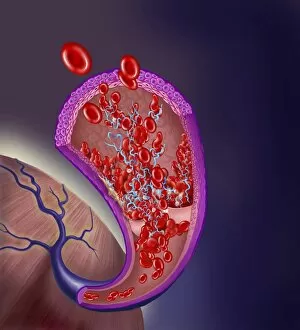
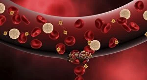
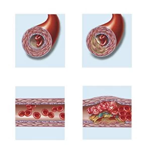
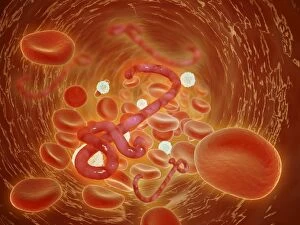
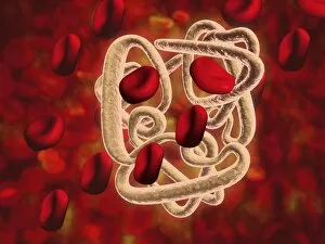
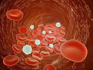
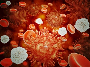
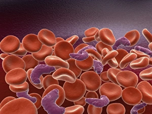
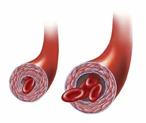
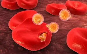
Hematology, the study of blood and its components, is a fascinating field that delves into the intricate workings of our circulatory system. Through advanced imaging techniques such as scanning electron microscopy (SEM), scientists have been able to capture mesmerizing images of red and white blood cells in action. In one captivating SEM image, we witness the vibrant dance between red and white blood cells. The crimson hue of red blood cells contrasts beautifully with their pale counterparts, showcasing the delicate balance required for optimal health. Another striking artwork showcases computer-generated imagery of red blood cells, highlighting their unique structure and function within our bodies. These tiny carriers of oxygen are essential for sustaining life itself. Haematopoietic stem cells take center stage in yet another SEM image. These remarkable cells possess the extraordinary ability to differentiate into various types of blood cells, ensuring a constant supply throughout our lifetime. The importance becomes even more evident when we observe a tuberculosis patient receiving a direct blood transfusion. This life-saving procedure relies on meticulous analysis by hematologists to match compatible donors with recipients accurately. One cannot discuss hematology without mentioning Paul Ehrlich, a pioneering German physician whose groundbreaking work revolutionized this field. His contributions paved the way for advancements in diagnosing and treating various hematological disorders. Histopathology plays an integral role in understanding pathophysiology related to conditions like diabetic foot ulcers. By examining tissue samples under a microscope, experts gain valuable insights into these complex wounds' underlying causes. A conceptual image featuring platelets alongside red and white blood cells symbolizes their collective effort in maintaining proper clotting mechanisms within our bloodstream. This harmonious collaboration ensures swift healing whenever injuries occur. Microscopic views offer us glimpses into diseases like leukemia through detailed images capturing abnormal leukemia cells infiltrating healthy tissues. Such visuals aid researchers in developing targeted therapies against this devastating condition. SEM images continue to captivate us with their portrayal of white blood cells and platelets.