
Hernia Collection
"Hernia: A Journey through Surgical Innovation and Healing" Step into the world surgery, where surgical instruments become the heroes in removing foreign bodies


All Professionally Made to Order for Quick Shipping
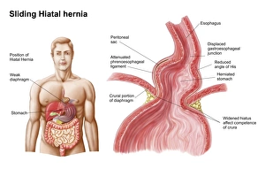
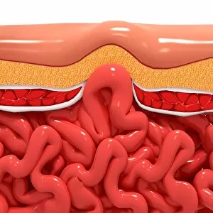
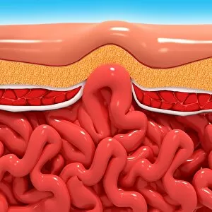
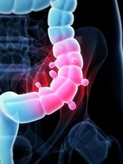
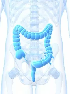
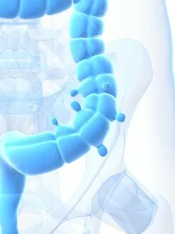
"Hernia: A Journey through Surgical Innovation and Healing" Step into the world surgery, where surgical instruments become the heroes in removing foreign bodies. From ancient times to modern innovations, these tools have played a crucial role in restoring health and well-being. In this captivating collection of images, we witness the evolution surgery. Coloured engravings from centuries ago depict intricate instruments specifically designed for this delicate procedure. These illustrations showcase not only the artistry but also the dedication of medical professionals striving to alleviate pain and discomfort. Amongst these historical depictions, we find a heartwarming lithograph showcasing an umbilical hernia in a young child. This reminder that even our little ones can face such challenges highlights the importance of early intervention and care. An advert from 1892 introduces us to Electropathic & Zander Institute, offering hope through innovative treatments for those suffering from hernias. It serves as a testament to human ingenuity and determination to find new solutions for complex medical conditions. The journey continues with glimpses into 18th-century surgical equipment – relics that paved the way for today's advanced techniques. These artifacts tell stories of skilled surgeons who braved uncharted territories in pursuit of healing their patients. A striking biomedical illustration reveals a hiatal hernia protruding into the thorax – an intricate depiction that showcases both anatomical knowledge and artistic prowess. Such visuals provide invaluable insights into how our bodies function and how they can be repaired when afflicted by this condition. Cross-section biomedical illustrations take us deeper into understanding hernias' development, repair processes, and incision sites necessary for successful treatment outcomes. The complexity behind each step is unveiled before our eyes, reminding us once again why medical professionals are true heroes in their field. Hernias may pose physical challenges, but they also represent resilience and triumph over adversity. Through continuous advancements in medicine, we gain greater insight into this condition, enabling us to provide effective care and support.