Fraud Blocker Skip to main content

Hormone Collection

"Hormone: The Intricate Symphony of the Body's Messengers" The medulla oblongata in the brain, a masterpiece of nature's artwork

Background imageHormone Collection: Medulla oblongata in the brain, artwork

Medulla oblongata in the brain, artwork
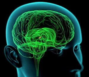
Hypothalamus in the brain. Computer artwork of a persons head showing the brain inside. The highlighted area shows the hypothalamus

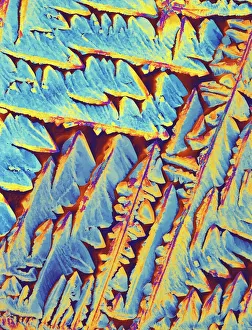
Background imageHormone Collection: Oxytocin hormone crystals, PLM C016 / 7196

Oxytocin hormone crystals, PLM C016 / 7196
Oxytocin. Polarised light micrograph (PLM) of crystals of the female hormone oxytocin. In women this hormone is secreted naturally by the pituitary gland

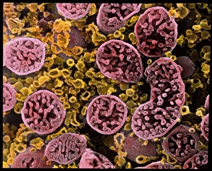
Background imageHormone Collection: Coloured SEM of mitochondria in ovarian cells

Coloured SEM of mitochondria in ovarian cells
Mitochondria. Coloured Scanning Electron Micro- graph (SEM) of mitochondria and smooth endoplasmic reticulum in an ovarian granulosa- lutein cell

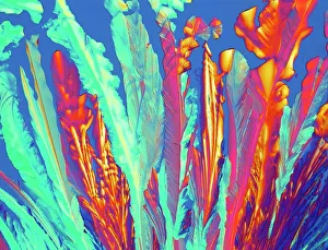
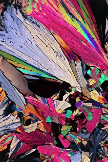
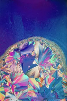
Background imageHormone Collection: Oxytocin crystals, light micrograph

Oxytocin crystals, light micrograph
Oxytocin. Polarised light micrograph of crystals of the female hormone oxytocin. In women this hormone is secreted naturally by the pituitary gland

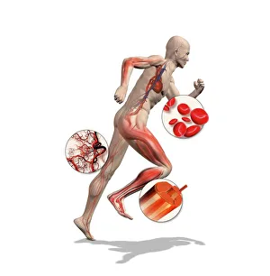
Background imageHormone Collection: Athlete physiology, artwork

Athlete physiology, artwork
Athlete physiology. Computer artwork of a male athlete, with enlargements to show the important physiological enhancements typical of most athletes

Background imageHormone Collection: Insulin molecule, artwork

Insulin molecule, artwork
Artwork of a molecule of human insulin, a hormone produced by the pancreas which controls levels of glucose in the blood

Background imageHormone Collection: Pancreas anatomy, artwork

Pancreas anatomy, artwork
Pancreas anatomy. Cut-away computer artwork of the pancreas, which secretes digestive enzymes and the hormones insulin and glucagon, which regulate blood sugar levels

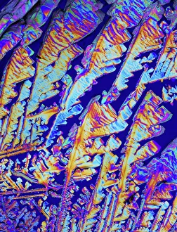
Background imageHormone Collection: PLM of crystals of testosterone

PLM of crystals of testosterone
^BMale sex hormone.^b Polarised light micrograph of crystals of testosterone

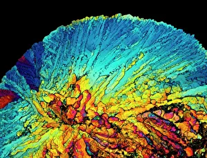
Background imageHormone Collection: Insulin crystals, light micrograph C017 / 8249

Insulin crystals, light micrograph C017 / 8249
Insulin. Polarised light micrograph (PLM) of crystals of the hormone insulin

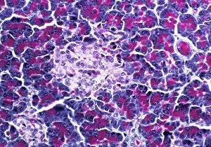
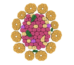
Background imageHormone Collection: Pancreatic islet of Langerhans

Pancreatic islet of Langerhans
Islet of Langerhans. Light micrograph of a section through an islet of Langerhans (pale, centre) in pancreas tissue

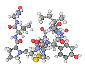
Background imageHormone Collection: Oxytocin neurotransmitter molecule

Oxytocin neurotransmitter molecule. Computer model showing the structure of the neurotransmitter and hormone Oxytocin

Background imageHormone Collection: Oxytocin hormone crystals, LM C016 / 7195

Oxytocin hormone crystals, LM C016 / 7195
Oxytocin. Polarised light micrograph of crystals of the female hormone oxytocin. In women this hormone is secreted naturally by the pituitary gland

Background imageHormone Collection: Thyroid anatomy, artwork C013 / 4675

Thyroid anatomy, artwork C013 / 4675
Thyroid anatomy. Computer artwork of the thyroid glands (pink) of the neck, shown in relation to the bodys bones and throat structures (white)

Background imageHormone Collection: Leptin, molecular model

Leptin, molecular model. This hormone is produced by adipose (fat) tissue. It interacts with receptors in the brains hypothalamus to signal when a person is full

Background imageHormone Collection: Progesterone hormone

Progesterone hormone
Polarised light micrograph of crystals of progesterone. X 40

Background imageHormone Collection: Fentanyl molecular structure. Fentanyl skeletal chemical formula

Fentanyl molecular structure. Fentanyl skeletal chemical formula. Chemical molecular formula vector illustration Fentanyl molecular structure. Fentanyl skeletal chemical formula

Background imageHormone Collection: Diagnosis of thyroid disease vector illustration

Diagnosis of thyroid disease vector illustration

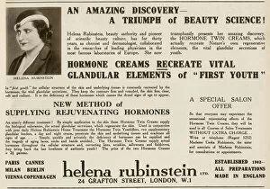
Background imageHormone Collection: Helena Rubinstein advertisement

Helena Rubinstein advertisement
Advertisement for Helena Rubinstein of Grafton Street, London, specifically hormone creams claiming to recreate vital glandular elements of " first youth". Date: 1932

Background imageHormone Collection: Kamov Ka-25

Kamov Ka-25
Russian Navy Kamov Ka-25 Helicopter Flying Date: 1976

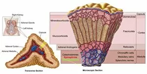
Background imageHormone Collection: Anatomy of adrenal gland, cross section

Anatomy of adrenal gland, cross section

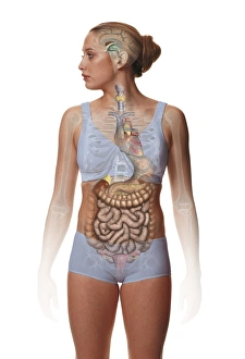
Background imageHormone Collection: Woman standing, body facing forward head turned to one side

Woman standing, body facing forward head turned to one side, illustration overlay showing skeleton and inner organs

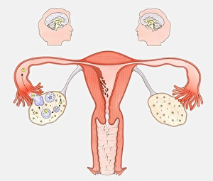
Background imageHormone Collection: Diagram showing the interaction between female sexual organs and the brain

Diagram showing the interaction between female sexual organs and the brain, on one side, the normal reproductive cycle, and on the other, the effect of the contraceptive pill

Background imageHormone Collection: Cretin aged 15. Cretinism in infants and children is caused by defective thyroid gland

Cretin aged 15. Cretinism in infants and children is caused by defective thyroid gland and failure to produce the hormone thyroxine. Sufferers are mentally and physically retarded

Background imageHormone Collection: Illustration of pituitary gland releasing hormones

Illustration of pituitary gland releasing hormones

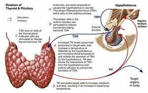
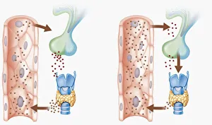
Background imageHormone Collection: Relation of thyroid and pituitary gland

Relation of thyroid and pituitary gland

Background imageHormone Collection: Emu All Emotional

Emu All Emotional
Overexcited Australian Emu (Dromaius novaehollandiae) hollering, bill wide open

Background imageHormone Collection: Female endocrine glands, artwork C016 / 7522

Female endocrine glands, artwork C016 / 7522
Female endocrine glands. Computer artwork showing the location of the endocrine glands (yellow) in a womans body

Background imageHormone Collection: Enteroendocrine cell, TEM

Enteroendocrine cell, TEM
Enteroendocrine cell. Transmission electron micrograph (TEM) of a section through an enteroendocrine cell within a gland of the stomach

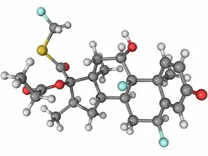
Background imageHormone Collection: Fluticasone asthma drug molecule

Fluticasone asthma drug molecule
Fluticasone, molecular model. This corticosteroid is used to treat asthma attacks

Background imageHormone Collection: Endocrine system, artwork

Endocrine system, artwork
Endocrine system. Computer artwork showing the locations of hormone-producing glands (endocrine glands) in the human body

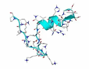
Background imageHormone Collection: Parathyroid hormone molecule

Parathyroid hormone molecule. Computer model showing the structure of parathyroid hormone (PTH), or parathormone

Background imageHormone Collection: Ghrelin hormone molecule

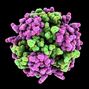
Ghrelin hormone molecule. Computer model showing the crystal structure of the human hormone ghrelin

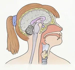
Background imageHormone Collection: Illustration showing connection of thyroid gland and pituitary gland to brain of pre-adolescent girl

Illustration showing connection of thyroid gland and pituitary gland to brain of pre-adolescent girl

Background imageHormone Collection: Digital cross section illustration of hypothalamus triggering chain reaction of reduced hormone production resulting in

Digital cross section illustration of hypothalamus triggering chain reaction of reduced hormone production resulting in falling glucose levels

Background imageHormone Collection: Cross section biomedical illustration of man endocrine system

Cross section biomedical illustration of man endocrine system

Background imageHormone Collection: Conceptual image of pancreatic islet of Langerhans

Conceptual image of pancreatic islet of Langerhans

Background imageHormone Collection: Pathway of a pain message via sensory nerve in injured muscle

Pathway of a pain message via sensory nerve in injured muscle, to pain gate in spinal cord to limbic system, frontal cortex and sensory cortex in the human brain

Background imageHormone Collection: Close-up view of thyroid gland

Close-up view of thyroid gland

Background imageHormone Collection: Schematic of the hypothalamus receiving nerve impulses from the body

Schematic of the hypothalamus receiving nerve impulses from the body and sending messages to the circulatory and nervous system

Background imageHormone Collection: Endorphins released in the hypothalamus travel down the spinal cord

Endorphins released in the hypothalamus travel down the spinal cord
Serotonin released in the brain travels down the spinal cord to close the pain gates and block pain messages

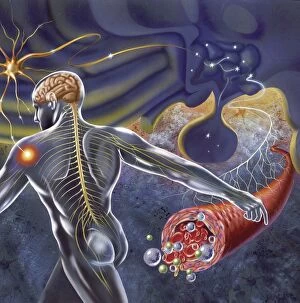
Background imageHormone Collection: Components of blood vessel interact with intracellular fluid

Components of blood vessel interact with intracellular fluid; white blood cells, red blood cells, oxygen, platelets and hormones

Background imageHormone Collection: Charles Herbert Best (1899-1978) Canadian physiologist: assisted Banting in isolating

Charles Herbert Best (1899-1978) Canadian physiologist: assisted Banting in isolating the hormone Insulin. Discovered the vitamin choline and the enzyme histaminase

Background imageHormone Collection: Illustration of hypophysis, or pituitary gland

Illustration of hypophysis, or pituitary gland
Medicine: Human anatomy, hypophysis, or pituitary gland. Drawing

Background imageHormone Collection: Vaccinium corymbosum (Blueberry)

Vaccinium corymbosum (Blueberry), trimming base of cutting just below a node and removing leaves from lower third of cutting, dish of hormone rooting powder nearby

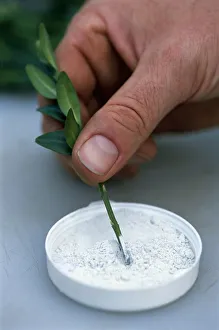
Background imageHormone Collection: Man dipping softwood leaf stem in hormone rooting compound

Man dipping softwood leaf stem in hormone rooting compound

Background imageHormone Collection: Person dipping cuttings in hormone rooting powder, close-up

Person dipping cuttings in hormone rooting powder, close-up

Background imageHormone Collection: Hands dusting incision on shoot of climbing plant with hormone rooting powder

Hands dusting incision on shoot of climbing plant with hormone rooting powder, using a paintbrush (propagation by simple layering), close-up

Background imageHormone Collection: Hand dipping bottom of evergreen cutting into hormone rooting powder, close-up

Hand dipping bottom of evergreen cutting into hormone rooting powder, close-up

Background imageHormone Collection: Contraceptive implant, X-ray C017 / 7390

Contraceptive implant, X-ray C017 / 7390
Contraceptive implant. X-ray of the arm of a 19 year old female showing a contraceptive implant (thin rod at upper centre)

Background imageHormone Collection: Tablet computer, insulin molecule F006 / 6311

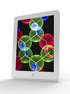
Tablet computer, insulin molecule F006 / 6311
Tablet computer showing a part of the molecule of human insulin. A single insulin molecule is made up of two chains of amino acids, the A and B chains, which are held together by di-sulphide bridges

Background imageHormone Collection: Insulin, artwork F007 / 7233

Insulin, artwork F007 / 7233
Insulin, computer artwork

Background imageHormone Collection: Insulin, artwork F007 / 7234

Insulin, artwork F007 / 7234
Insulin, computer artwork

Background imageHormone Collection: Human serum albumin molecule F007 / 9904

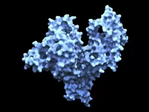
Human serum albumin molecule F007 / 9904
Human serum albumin, molecular model. Albumin is the most abundant protein in human blood plasma. One of albumins functions is to transport fatty acids to the liver

Background imageHormone Collection: Human serum albumin molecule F007 / 9905

Human serum albumin molecule F007 / 9905
Human serum albumin, molecular model. Albumin is the most abundant protein in human blood plasma. One of albumins functions is to transport fatty acids to the liver

Background imageHormone Collection: Hydrocortisone ointment F008 / 3400

Hydrocortisone ointment F008 / 3400
Hydrocortisone ointment in tube

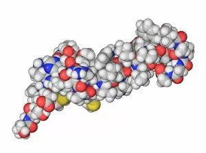
Background imageHormone Collection: Insulin A chain molecule

Insulin A chain molecule
Insulin A chain. Computer model of an A chain of human insulin that has been synthesized on a crosslinked polystyrene solid support. This is an example of solid phase peptide synthesis (SPPS)

Background imageHormone Collection: Noradrenaline norepinephrine molecule

Noradrenaline norepinephrine molecule
Noradrenaline (norepinephrine), belonging to the group of catecholamines, molecular model. Catecholamine acting also as a hormone and a neurotransmitter

Background imageHormone Collection: Adrenaline epinephrine molecule

Adrenaline epinephrine molecule
Adrenaline (epinephrine) is a hormone and a neurotransmitter. It is used to treat a number of conditions like cardiac arrest, anaphylaxis, and superficial bleeding

Background imageHormone Collection: Lixivaptan hyponatremia drug F007 / 0159

Lixivaptan hyponatremia drug F007 / 0159
Lixivaptan hyponatremia drug, molecular model. Lixivaptan blocks vasopressin from binding the vasopressin 2 receptor, and is used to treat heart failure in hyponatremia patients

Background imageHormone Collection: Lixivaptan hyponatremia drug F007 / 0158

Lixivaptan hyponatremia drug F007 / 0158
Lixivaptan hyponatremia drug, molecular model. Lixivaptan blocks vasopressin from binding the vasopressin 2 receptor, and is used to treat heart failure in hyponatremia patients

Background imageHormone Collection: Budesonide steroid drug F007 / 0117

Budesonide steroid drug F007 / 0117
Budesonide steroid drug, molecular model. Atoms are represented as spheres and are colour-coded: hydrogen (white), carbon (grey) and oxygen (red)


All products are expertly crafted, using premium materials, tailored to your specifications and promptly shipped

"Hormone: The Intricate Symphony of the Body's Messengers" The medulla oblongata in the brain, a masterpiece of nature's artwork, orchestrates an intricate dance of hormones. Among them, oxytocin hormone crystals shimmer like precious gems under the microscope (PLM C016 / 7196), revealing their profound role in human connection and bonding. Delving deeper into cellular realms, a colored SEM image showcases mitochondria within ovarian cells, highlighting their crucial involvement in hormone production. Meanwhile, light micrographs capture oxytocin crystals (LM C016 / 7195) and insulin crystals (C017 / 8249), unveiling their mesmerizing structures that hold vital functions within our bodies. Exploring athlete physiology through captivating artwork unveils the remarkable impact hormones have on performance and endurance. These chemical messengers drive athletes to push beyond limits and achieve greatness. Artistic depictions also shed light on the complex world of molecular biology. An intricately designed insulin molecule stands as a testament to its pivotal role in regulating blood sugar levels while offering hope for those with diabetes. The pancreas anatomy comes alive through vibrant illustrations showcasing its significance as both an endocrine and digestive organ. It secretes essential hormones such as insulin that keep our bodies functioning optimally. Testosterone takes center stage with PLM capturing crystal formations that symbolize strength and masculinity. This hormone plays a fundamental role not only in physical development but also influences mood, cognition, and overall well-being. Returning to oxytocin - known as the "love hormone" - we delve into its dual identity as both neurotransmitter and hormonal messenger. Its delicate crystalline structure reveals itself once again under microscopic scrutiny (Oxytocin hormone crystals). Lastly, an artistic portrayal of thyroid anatomy reminds us how this small gland holds immense power over metabolism regulation (artwork C013 / 4675). Hormones secreted by this butterfly-shaped organ impact our energy levels, weight management, and overall health.

© Copyright Media Storehouse, All Rights Reserved 2003 - 2024
Facebook     Pinterest     100% Payment Secure     Reviews IO