
Human Knee Collection
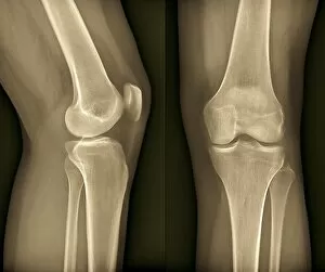
"The Human Knee: A Complex Joint Revealed Through X-rays" Normal knees


All Professionally Made to Order for Quick Shipping
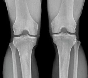
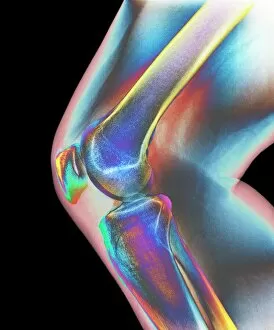
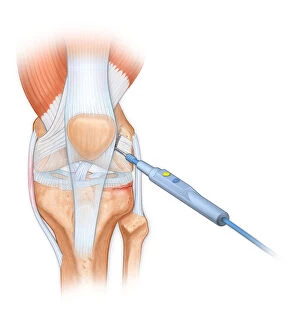
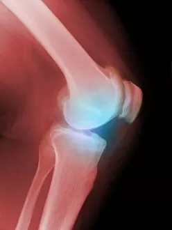
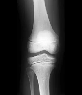
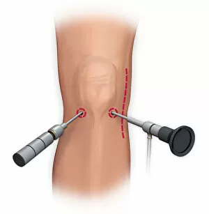
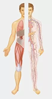
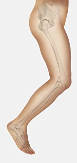
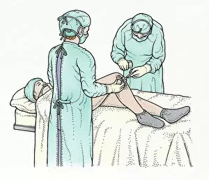
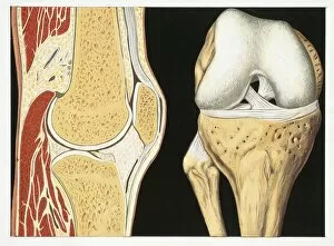
"The Human Knee: A Complex Joint Revealed Through X-rays" Normal knees, X-ray: Peek into the intricate structure of a healthy knee joint through this fascinating X-ray image. Healthy knee, X-ray: Witness the beauty of a perfectly functioning knee captured in this remarkable X-ray. Teen girl with vinyl records and record player, sitting on floor: As she enjoys her music collection, let's appreciate our knees that allow us to sit comfortably and indulge in our favorite activities. Knee joint: Delve into the inner workings of the incredible knee joint - a marvel of engineering enabling movement and stability. Total knee replacement, X-rays: Discover how medical advancements restore mobility as we explore these enlightening X-rays showcasing total knee replacements. Arthritic knees, X-ray: Uncover the impact of arthritis on knees through this revealing X-ray image - a reminder to cherish our joints' health. Bovie used to cut through retinaculum and clean up femur of Displaced patellar knee: Witness surgical precision at work as doctors utilize advanced techniques to address complex knee injuries effectively. Illustration of the anterior knee, articular surface meniscus: Gain insight into the delicate structures within your anterior knee region with this detailed illustration highlighting articular surfaces and menisci. Arthritic knee, X-ray: Explore an arthritic condition affecting one's quality of life by examining this illuminating x-ray image capturing its effects on the human body. Normal child's knee, x-ray : Marvel at nature's perfection as you observe an innocent child's pristine x-rayed kneecap – reminding us all about preserving their well-being for years to come. A leg and kneecap injury sustained from repetitive motion: Reflect upon the consequences that repetitive motions can have on our bodies as we contemplate this unfortunate leg and kneecap injury, urging us to prioritize self-care and ergonomics.