Fraud Blocker Skip to main content

Human Spine Collection

"The Human Spine: A Marvel of Structure and Support" The human spine, also known as the backbone

Background imageHuman Spine Collection: Human Anatomy - Backbone including Ribs and Pelvis

Human Anatomy - Backbone including Ribs and Pelvis
Vintage engraving of the human Backbone including Ribs and Pelvis. 19th Century

Background imageHuman Spine Collection: Human Anatomy Skeleton and muscles of the body

Human Anatomy Skeleton and muscles of the body
Vintage engraving of Human Anatomy, the bones of the skeleton and muscles of the body, 1898

Background imageHuman Spine Collection: Diagram of the human spine, side view

Diagram of the human spine, side view

Background imageHuman Spine Collection: Human spine anatomy illustration 1866

Human spine anatomy illustration 1866
Atlas d anatomie descriptive du corps humain

Background imageHuman Spine Collection: Digital illustration of human nervous system connected to spinal cord and brain

Digital illustration of human nervous system connected to spinal cord and brain

Background imageHuman Spine Collection: Human spine, computer artwork

Human spine, computer artwork
Computer artwork showing the human spine emphasizing the lumbar vertebrae. The sacrum is just visible in the lower left and the skull in the upper right corner of the image

Background imageHuman Spine Collection: Dorsal vertebrae anatomy illustration 1866

Dorsal vertebrae anatomy illustration 1866
Atlas d anatomie descriptive du corps humain

Background imageHuman Spine Collection: Cervical vertebrae anatomy illustration 1866

Cervical vertebrae anatomy illustration 1866
Atlas d anatomie descriptive du corps humain C. Bonamy - Paul Broca Victor Masson et Fils Paris 1866

Background imageHuman Spine Collection: Anterior cross section of spinal column revealing how vertebra fits round spinal cord

Anterior cross section of spinal column revealing how vertebra fits round spinal cord

Background imageHuman Spine Collection: Rear view of human skeletal system showing upper back

Rear view of human skeletal system showing upper back

Background imageHuman Spine Collection: Visualization of human diaphragm

Visualization of human diaphragm

Background imageHuman Spine Collection: Medical chart showing the signs and symptoms of sciatica

Medical chart showing the signs and symptoms of sciatica

Background imageHuman Spine Collection: Human skeleton and a skeleton of a monkey, anatomical illustration

Human skeleton and a skeleton of a monkey, anatomical illustration

Background imageHuman Spine Collection: The human skeleton engraving 1895

The human skeleton engraving 1895
The human skeleton engraving

Background imageHuman Spine Collection: Diagram of a human thoracic vertebrae including the intervertebral disk

Diagram of a human thoracic vertebrae including the intervertebral disk, vertebral process and ligaments

Background imageHuman Spine Collection: Man sitting on floor with one hand behind head, other hand on his knee, looking up

Man sitting on floor with one hand behind head, other hand on his knee, looking up, illustration of his skeleton overlaid

Background imageHuman Spine Collection: Illustration of human Central Nervous System showing brain, spinal cord, veins and arteries

Illustration of human Central Nervous System showing brain, spinal cord, veins and arteries

Background imageHuman Spine Collection: Digital illustration of brain connected to spinal nerves branching out from spinal cord

Digital illustration of brain connected to spinal nerves branching out from spinal cord

Background imageHuman Spine Collection: 3D rendering of human vertebral column with skull

3D rendering of human vertebral column with skull

Background imageHuman Spine Collection: Three dimensional view of female sternum and rib cage

Three dimensional view of female sternum and rib cage

Background imageHuman Spine Collection: Human spine, side view

Human spine, side view

Background imageHuman Spine Collection: Anatomy of human pelvic bone

Anatomy of human pelvic bone

Background imageHuman Spine Collection: The human skeleton and muscular system, front view

The human skeleton and muscular system, front view
Medical illustration of the human skeleton and muscular system, front view

Background imageHuman Spine Collection: Muscles of the back and neck

Muscles of the back and neck (splenius capitis muscle, trapezius muscle)

Background imageHuman Spine Collection: Digital illustration of pain signal to area of somatosensory cortex in human brain via spine

Digital illustration of pain signal to area of somatosensory cortex in human brain via spine

Background imageHuman Spine Collection: Spine and Ligaments anatomy illustration 1866

Spine and Ligaments anatomy illustration 1866
Atlas d anatomie descriptive du corps humain C. Bonamy - Paul Broca Victor Masson et Fils Paris 1866

Background imageHuman Spine Collection: Back muscles

Back muscles
Antique illustration of a back muscles

Background imageHuman Spine Collection: Back muscles anatomy

Back muscles anatomy
Antique illustration of a back muscles

Background imageHuman Spine Collection: Section diagram depicting the human vertebral column, spinal nerve

Section diagram depicting the human vertebral column, spinal nerve, spinal cord and vertebra of the delicate spina

Background imageHuman Spine Collection: Illustration showing human spine and skull

Illustration showing human spine and skull

Background imageHuman Spine Collection: Illustration of bundle of nerves in human spine

Illustration of bundle of nerves in human spine

Background imageHuman Spine Collection: Digital illustration of human spine with damaged area highlighted in blue resulting in quadriplegia

Digital illustration of human spine with damaged area highlighted in blue resulting in quadriplegia

Background imageHuman Spine Collection: Cross section digital illustration of spinal nerve fibres and convey motor signals highlighted in

Cross section digital illustration of spinal nerve fibres and convey motor signals highlighted in pink and green

Background imageHuman Spine Collection: Digital illustration of woman with head in profile showing brain connected to spinal cord

Digital illustration of woman with head in profile showing brain connected to spinal cord

Background imageHuman Spine Collection: Digital illustration of of human nervous system

Digital illustration of of human nervous system

Background imageHuman Spine Collection: Digital illustration of human brain stem with cerebellum removed revealing medulla and axons

Digital illustration of human brain stem with cerebellum removed revealing medulla and axons

Background imageHuman Spine Collection: Digital illustration of head in profile showing skull, brain, and spine

Digital illustration of head in profile showing skull, brain, and spine

Background imageHuman Spine Collection: Digital illustration of human brain with spine highlighted in pink

Digital illustration of human brain with spine highlighted in pink

Background imageHuman Spine Collection: Illustration of doctor pointing at x-ray of pelvis and spine

Illustration of doctor pointing at x-ray of pelvis and spine

Background imageHuman Spine Collection: Cross section biomedical illustration of epidural anaesthesia during labour

Cross section biomedical illustration of epidural anaesthesia during labour

Background imageHuman Spine Collection: Cross section biomedical illustration of the brain and skull at 18 years of age

Cross section biomedical illustration of the brain and skull at 18 years of age

Background imageHuman Spine Collection: Cross section biomedical illustration of voluntary pathway of human brain

Cross section biomedical illustration of voluntary pathway of human brain

Background imageHuman Spine Collection: Cross section biomedical illustration of spinal reflex pathway of human brain

Cross section biomedical illustration of spinal reflex pathway of human brain

Background imageHuman Spine Collection: Cross section biomedical illustration of site of incision for microdiscectomy surgical procedure

Cross section biomedical illustration of site of incision for microdiscectomy surgical procedure

Background imageHuman Spine Collection: Rear view of human skeleton showing kidney and nervous system

Rear view of human skeleton showing kidney and nervous system

Background imageHuman Spine Collection: Human skeletal system

Human skeletal system

Background imageHuman Spine Collection: Medical illustration of a womans skull and skeletal system

Medical illustration of a womans skull and skeletal system, three dimensional view


All Professionally Made to Order for Quick Shipping

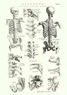
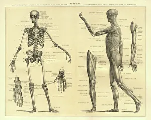
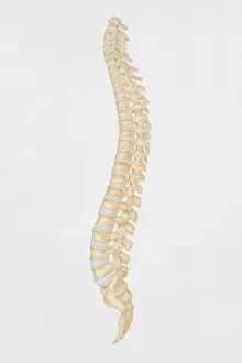
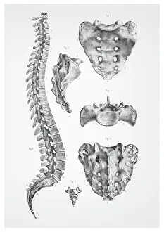
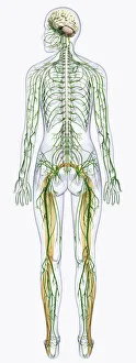
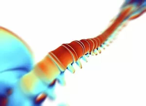
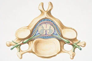
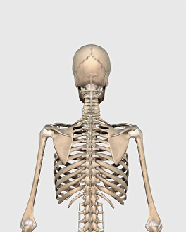
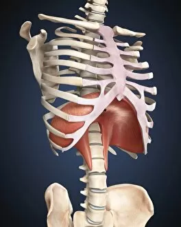
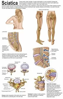
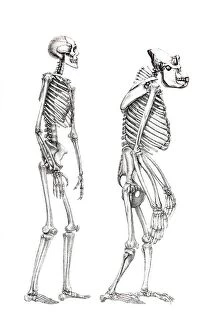
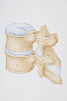
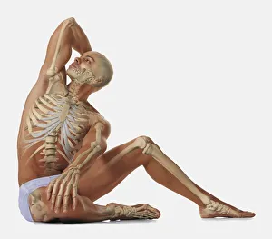
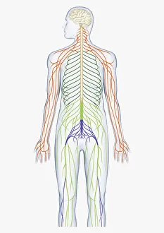
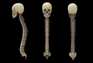
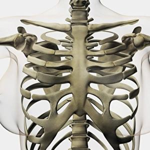
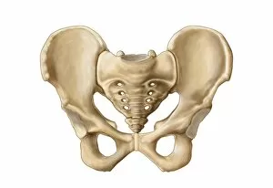
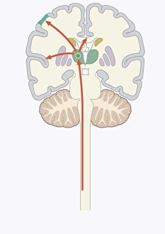
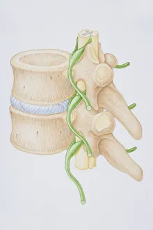
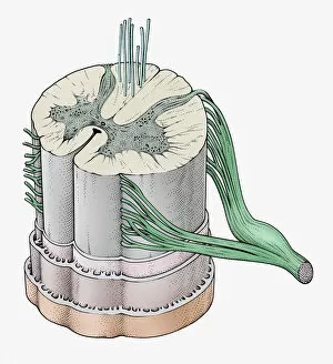
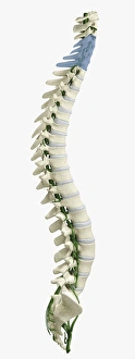
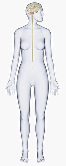
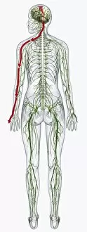
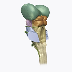
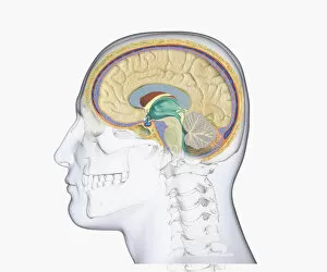
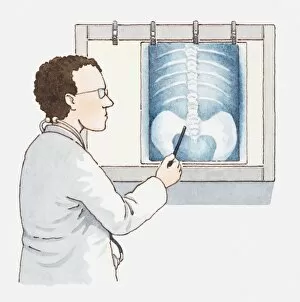
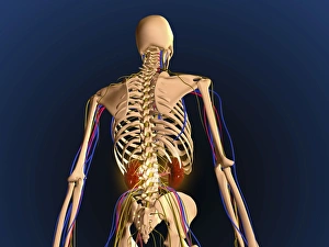
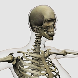
"The Human Spine: A Marvel of Structure and Support" The human spine, also known as the backbone, is a remarkable anatomical feature that provides stability and flexibility to our bodies. Comprising vertebrae, ribs, and the pelvis, it forms the central axis of our skeletal system. Intricate illustrations from 1866 showcase the detailed anatomy of this vital structure. From a side view diagram to dorsal and cervical vertebrae illustrations, these depictions offer insights into its complexity. The digital artwork further highlights how the nervous system connects with the spinal cord and brain—a testament to its role in transmitting signals throughout our body. Computer-generated imagery brings forth an artistic representation of this intricate network—the human spine—showcasing its significance in maintaining posture and facilitating movement. It serves as a reminder of how technology can enhance our understanding of biological structures. Moreover, when viewed from behind, we gain a glimpse into the upper back's skeletal framework—an area where muscles intertwine with bones to provide support for everyday activities. Additionally, visualizing the diaphragm sheds light on its relationship with respiration—a process intricately linked to spinal alignment. Anatomical comparisons between humans and monkeys reveal intriguing similarities in their skeletons—an evolutionary connection that underscores shared characteristics within different species. Lastly, an engraving depicting the cardiovascular system reminds us that while focusing on individual structures like the spine is crucial; it is equally important to acknowledge their interconnectedness within our bodies' complex systems. Exploring various aspects anatomy—from historical illustrations to modern renderings—allows us to appreciate both its structural elegance and functional importance. Understanding this integral part of ourselves fosters awareness about maintaining good spinal health for overall well-being.