
Iliac Collection
The iliac region, a vital part of the human anatomy, encompasses various structures and systems that play crucial roles in our overall health


All Professionally Made to Order for Quick Shipping
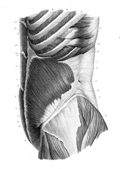
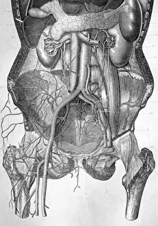
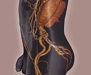
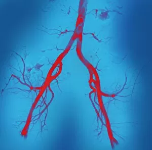
The iliac region, a vital part of the human anatomy, encompasses various structures and systems that play crucial roles in our overall health. From abdominal arteries to nerve plexuses, this area holds significant importance. In an X-ray labeled P206 / 0309, we can observe the intricate network of abdominal arteries coursing through the iliac region. These blood vessels supply oxygenated blood to essential organs within the abdomen. An engraving from 1866 showcases frontal trunk anatomy, providing a detailed view of the iliac region's internal structures. This illustration serves as a valuable resource for medical professionals studying this complex area. A medical illustration depicting both male lymphatic system and reproductive organs highlights their proximity in the iliac region. It emphasizes how these systems intertwine and function together harmoniously. Similarly, another artwork portrays a female body in dynamic posture with superimposed lymphatic system. This visual representation aids in understanding how lymphatic vessels traverse through the iliac region while maintaining balance within her body. An 1880 artwork labeled C017 / 6913 delves into deep abdominal organs found within the iliac region. This piece provides insights into their placement and relationship with surrounding structures. Atherosclerosis is visually captured through a remarkable 3D CT scan image. The depiction reveals how this condition affects arterial walls within the iliac region, emphasizing its impact on blood flow dynamics. Artworks C016 / 6808 and C016 / 6809 focus on left hip and right hip respectively along with their corresponding nerve plexuses present in the iliac area. These illustrations help comprehend neural pathways responsible for sensory-motor functions related to lower limbs originating from these regions. Total mesorectal excision is showcased via artwork C016 /6996 which demonstrates surgical intervention techniques performed within or near the iliac area for rectal cancer treatment purposes. An exquisite artwork dating back to1825 captures abdominal lymph vessels, shedding light on their intricate network within the iliac region.