Fraud Blocker Skip to main content

Immunology Collection (page 7)

"Unleashing the Power of Immunology: Exploring the Intricate World of Immune Responses" Immunology

Background imageImmunology Collection: Activated plasma cell, TEM

Activated plasma cell, TEM
Activated plasma cell, coloured transmission electron micrograph (TEM). Plasma cells are derived from stimulated B lymphocytes (white blood cells)

Background imageImmunology Collection: Basophil white blood cell, TEM

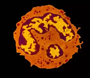
Basophil white blood cell, TEM
Basophil white blood cell. Transmission electron micrograph (TEM) of section through a circulating basophil cell. Basophils are the smallest and least common of the white blood cells

Background imageImmunology Collection: Lymphoblastic sarcoma, light micrograph

Lymphoblastic sarcoma, light micrograph
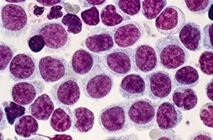
Lymphoblastic sarcoma. Light micrograph of blood cells from bone marrow in a case of lymphoblastic sarcoma. These small lymphoblasts (dark red) characteristically have conspicuous nucleoli

Background imageImmunology Collection: Plasmocyte blood cell, light micrograph

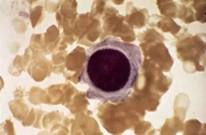
Plasmocyte blood cell, light micrograph. Plasmocytes, also called plasma cells, are derived from stimulated B lymphocytes (white blood cells)

Background imageImmunology Collection: Multiple myeloma, light micrograph

Multiple myeloma, light micrograph
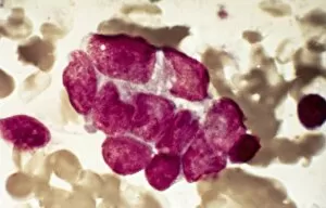
Multiple myeloma. Light micrograph of dystrophy (abnormality) of plasmocyte blood cells from bone marrow in a case of multiple myeloma, also known as Kahlers disease or plasma cell myeloma

Background imageImmunology Collection: Lymphoplasmacytic lymphoma, micrograph

Lymphoplasmacytic lymphoma, micrograph
Lymphoplasmacytic lymphoma. Light micrograph of a mixture of small lymphocytes and plasma cells from bone marrow in a case of lymphoplasmacytic lymphoma, also known as Waldenstroms macroglobulinemia

Background imageImmunology Collection: Hairy cell leukaemia, light micrograph

Hairy cell leukaemia, light micrograph
Hairy cell leukaemia. Light micrograph of blood cells in a case of hairy cell leukaemia. This sample, treated with May-Grunwald-Giemsa (MGG) stain

Background imageImmunology Collection: Mast cells, light micrograph

Mast cells, light micrograph
Mast cells. Light micrograph of several mast cells (pink rounded objects). These leucocytes (white blood cells) contain the chemical mediators histamine, serotonin and heparin
Background imageImmunology Collection: Lymphocyte white blood cell, TEM

Lymphocyte white blood cell, TEM
Lymphocyte white blood cell. Transmission electron micrograph (TEM) of a section through lymphocyte, showing the nucleus (large, round, centre), endoplasmic reticulum (ER)

Background imageImmunology Collection: Acute lymphoblastic leukaemia, micrograph

Acute lymphoblastic leukaemia, micrograph
Acute lymphoblastic leukaemia. Light micrograph of blood cells from bone marrow in a case of acute lymphoblastic leukaemia. Leukaemia is a cancer where certain blood cells form in excess

Background imageImmunology Collection: Malignant histiocytosis, light micrograph

Malignant histiocytosis, light micrograph
Malignant histiocytosis. Light micrograph of blood cells in a case of malignant histiocytosis. Hystiocytosis is an excess number of histiocytes

Background imageImmunology Collection: Blood cells, light micrograph

Blood cells, light micrograph
Blood cells. Light micrograph of red blood cells (erythrocytes, light blue) and white blood cells (leucocytes, nuclei stained purple)

Background imageImmunology Collection: Chediak-Higashi syndrome, micrograph

Chediak-Higashi syndrome, micrograph
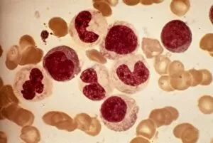
Chediak-Higashi syndrome. Light micrograph of blood cells in a case of Chediak-Higashi syndrome. The cells include neutrophil white blood cells with abnormal granules

Background imageImmunology Collection: Metastatic breast cancer, micrograph

Metastatic breast cancer, micrograph
Metastatic breast cancer. Light micrograph of blood cells in a case of metastatic (secondary) medullary breast cancer. Medullary breast cancer is a rare form of breast cancer that is diagnosed by

Background imageImmunology Collection: Metastatic lung cancer, micrograph

Metastatic lung cancer, micrograph
Metastatic lung cancer. Light micrograph of blood cells in a case of metastatic (secondary) medullary lung cancer. Medullary cancer (carcinoma)

Background imageImmunology Collection: Alders anomaly, light micrograph

Alders anomaly, light micrograph
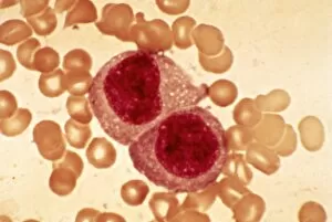
Alders anomaly. Light micrograph of an abnormal lymphocyte white blood cell (centre) from a blood sample in a case of Alders anomaly

Background imageImmunology Collection: Granulocyte white blood cell, TEM

Granulocyte white blood cell, TEM
Granulocyte white blood cell. Coloured transmission electron micrograph (TEM) of a neutrophil granulocyte, a type of white blood cell

Background imageImmunology Collection: Acute leukaemia, light micrograph

Acute leukaemia, light micrograph
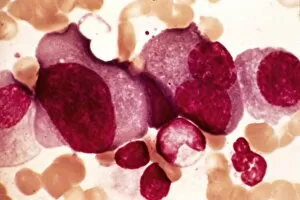
Acute leukaemia. Light micrograph of blood cells from bone marrow in a case of acute leukaemia. These cells include a polynuclear degranulated basophil and myeloblasts

Background imageImmunology Collection: Leukaemia cell, SEM

Leukaemia cell, SEM
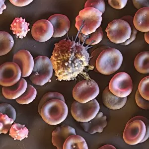
Leukaemia cell. Coloured scanning electron micrograph (SEM) of a leukaemic (cancerous) lymphocyte white blood cell (green), amongst normal red blood cells (erythrocytes, red)

Background imageImmunology Collection: Chronic myeloid leukaemia, micrograph

Chronic myeloid leukaemia, micrograph
Chronic myeloid leukaemia. Light micrograph of blood cells from bone marrow in a case of chronic myeloid leukaemia. Leukaemia is a cancer where certain blood cells form in excess

Background imageImmunology Collection: Granulocyte blood cells, light micrograph

Granulocyte blood cells, light micrograph
Granulocyte blood cells. Light micrograph of two types of granulocytes, a type of white blood cell (leucocyte). At left is a neutrophil, with an eosinophil at right

Background imageImmunology Collection: Langerhans cell histiocytosis, micrograph

Langerhans cell histiocytosis, micrograph
Langerhans cell histiocytosis. Light micrograph of blood cells in a case of Langerhans cell histiocytosis (LCH), also called X-type histiocytosis

Background imageImmunology Collection: Hodgkins disease, light micrograph

Hodgkins disease, light micrograph
Hodgkins disease. Light micrograph of two non-dystrophic histiocyte blood cells (centre) in a case of Hodgkins disease, also called Hodgkins lymphoma

Background imageImmunology Collection: Antibodies and B cells, artwork C016 / 6255

Antibodies and B cells, artwork C016 / 6255
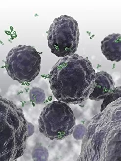
Antibodies and B cells. Computer artwork showing antibodies (green) neutralising cytokine activity in B cell (large, round) white blood cells

Background imageImmunology Collection: Antibodies and B cells, artwork C016 / 6254

Antibodies and B cells, artwork C016 / 6254
Antibodies and B cells. Computer artwork showing antibodies (green) neutralising cytokine activity in B cell (large, round) white blood cells

Background imageImmunology Collection: Blood cells, light micrograph C015 / 7130

Blood cells, light micrograph C015 / 7130
Blood cells. Light micrograph of red blood cells (erythrocytes, red) and white blood cells (leucocytes, nuclei stained pink)

Background imageImmunology Collection: Simian virus (SV40) large T antigen C015 / 7069

Simian virus (SV40) large T antigen C015 / 7069
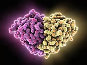
Simian virus (SV40) large T antigen, molecular model. This antigen is from the simian vacuolating virus 40 (SV40). Large T antigens play a role in regulating the viral life cycle of

Background imageImmunology Collection: Thymus gland cancer, light micrograph C015 / 7128

Thymus gland cancer, light micrograph C015 / 7128
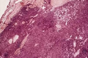
Thymus cancer. Light micrograph of a section through a thymus gland with thymoma cancer. The thymus is a gland in the upper chest that plays an important role in the immune system

Background imageImmunology Collection: Neutrophil white blood cell, illustration C018 / 0741

Neutrophil white blood cell, illustration C018 / 0741
Neutrophil white blood cell, illustration. Neutrophils are the most abundant white blood cell in humans. They have a multi-lobed nucleus (dark purple)

Background imageImmunology Collection: Blood cells, illustration C018 / 0802

Blood cells, illustration C018 / 0802
Blood cells. All cellular blood components originate from the same cell, the haematopoietic stem cell. The stem cell differentiates into two types of progenitor cells

Background imageImmunology Collection: Human immune response molecule complex

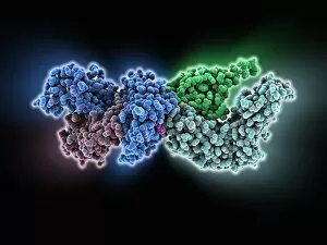
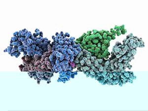
Human immune response molecule complex. Molecular model showing a human T-cell receptor and an HLA-A leukocyte (white blood cell) antigen bound to a TAX peptide from a virus

Background imageImmunology Collection: Microglial white blood cell, SEM C019 / 0247

Microglial white blood cell, SEM C019 / 0247
Microglial white blood cell, SEM

Background imageImmunology Collection: Microglial white blood cell, SEM C019 / 0246

Microglial white blood cell, SEM C019 / 0246
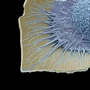
Microglia. Coloured scanning electron micrograph (SEM) of an activated microglial white blood cell. This immune system cell is found in the brain and spinal cord

Background imageImmunology Collection: Microglial white blood cell, SEM C019 / 0245

Microglial white blood cell, SEM C019 / 0245
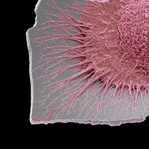
Microglia. Coloured scanning electron micrograph (SEM) of an activated microglial white blood cell. This immune system cell is found in the brain and spinal cord

Background imageImmunology Collection: Human immune response molecule complex C014 / 0871

Human immune response molecule complex C014 / 0871
Human immune response molecule complex. Molecular model showing a human T-cell receptor and an HLA-A leukocyte (white blood cell) antigen bound to a TAX peptide from a virus

Background imageImmunology Collection: Monocyte white blood cell, TEM

Monocyte white blood cell, TEM
Monocyte white blood cell. Transmission electron micrograph (TEM) of a section through a monocyte white blood cell, showing the cell nucleus (blue)

Background imageImmunology Collection: Virus, artwork C014 / 4951

Virus, artwork C014 / 4951
Computer artwork of a generic virus particle

Background imageImmunology Collection: Alemtuzumab antibody molecule

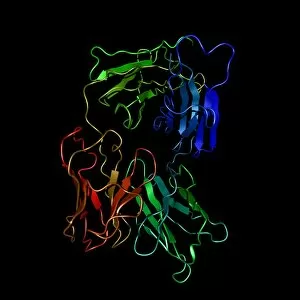
Alemtuzumab antibody molecule. Computer model showing the molecular structure of the monoclonal antibody and cancer drug alemtuzumab

Background imageImmunology Collection: Human lymphatic system, artwork

Human lymphatic system, artwork
Human lymphatic system. Artwork of a front view of a human, showing the lymphatic system (yellow). Also seen are associated organs including the spleen (centre right)

Background imageImmunology Collection: Alexander Fleming, caricature

Alexander Fleming, caricature
Alexander Fleming (1881-1955). Caricature of the Scottish biologist and pharmacologist Alexander Fleming holding a Petri dish

Background imageImmunology Collection: HIV invading white blood cell, artwork

HIV invading white blood cell, artwork
HIV invading white blood cell. Computer artwork showing HIV (human immunodeficiency virus) particles (virions, purple) invading a white blood cell (large, centre) in the human bloodstream

Background imageImmunology Collection: Blood cells, SEM C016 / 8026

Blood cells, SEM C016 / 8026
Blood cells. Coloured scanning electron micrograph (SEM) of a lymphocyte white blood cell (centre) and red blood cells (erythrocytes, circular)

Background imageImmunology Collection: Cat allergen protein, molecular model C015 / 3962

Cat allergen protein, molecular model C015 / 3962
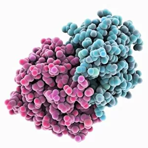
Cat allergen protein. Molecular model of the tetrameric form of the major cat allergen fel d 1 (Felis domesticus allergen 1)

Background imageImmunology Collection: Cat allergen protein, molecular model

Cat allergen protein, molecular model
Cat allergen protein. Molecular model of the tetrameric form of the major cat allergen fel d 1 (Felis domesticus allergen 1)

Background imageImmunology Collection: MHC protein-antigen complex

MHC protein-antigen complex. Computer model showing a histocompatibility antigen complexed to a class II MHC (major histocompatibility complex) protein molecule

Background imageImmunology Collection: Simian virus (SV40) large T antigen C015 / 7070

Simian virus (SV40) large T antigen C015 / 7070
Simian virus (SV40) large T antigen, molecular model. This antigen is from the simian vacuolating virus 40 (SV40). Large T antigens play a role in regulating the viral life cycle of

Background imageImmunology Collection: Immunoglobulin G antibody molecule C015 / 6789

Immunoglobulin G antibody molecule C015 / 6789
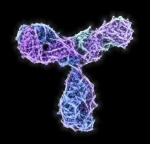
Immunoglobulin G antibody molecule. Computer artwork of a model of the secondary structure of immunoglobulin G (IgG). This is the most abundant immunoglobulin and is found in all body fluids

Background imageImmunology Collection: Activated macrophage, SEM C015 / 6377

Activated macrophage, SEM C015 / 6377
Activated macrophage. Coloured scanning electron micrograph (SEM) of an active macrophage white blood cell. Macrophages are cells of the bodys immune system that are found in the tissues rather than


All Professionally Made to Order for Quick Shipping

"Unleashing the Power of Immunology: Exploring the Intricate World of Immune Responses" Immunology, a fascinating field that delves into the complex mechanisms of our immune system, holds immense potential in combating diseases. T lymphocytes and cancer cells engage in a constant battle for supremacy, as depicted by SEM C001 / 1679. These tiny warriors play a crucial role in identifying and eliminating abnormal cells. The Immunoglobulin G antibody molecule (F007 / 9894) stands tall as one of our body's most powerful defenders against pathogens. Its remarkable structure enables it to neutralize harmful invaders with precision and efficiency. Neutrophils, exemplified by SEM C018 / 8596, showcase their extraordinary ability to engulf MRSA bacteria—an awe-inspiring sight indeed. Meanwhile, dendritic cells (artwork) act as vigilant sentinels, capturing antigens and presenting them to other immune cells for recognition. TEM reveals an up-close view of human white blood cells bearing HLA antigens—a key component in distinguishing self from non-self. Antibodies (artwork), resembling elegant warriors on a mission, bind specifically to foreign substances to mark them for destruction. HIV reverse transcription enzyme serves as a reminder of the challenges faced by immunologists worldwide. This relentless virus exploits our own cellular machinery but continues to be targeted through innovative research efforts. Human macrophages (TEM) demonstrate their exceptional phagocytic abilities while basophil white blood cells stand ready at the frontlines—both integral players in mounting effective immune responses against invading pathogens. Intriguingly captured by SEM imagery is bacteria infecting a macrophage—a visual representation highlighting how these microscopic organisms can exploit host defenses while also serving as valuable tools for studying infection dynamics. Lastly, Dohle bodies within blood cells offer insights into various pathological conditions affecting neutrophils—an essential clue guiding immunologists towards understanding and treating immune disorders.