
Implanted Collection
"Implanted: Exploring the Intricacies of Human Intervention" Step into a world where reality blurs with imagination, as we delve into the concept of being "implanted


All Professionally Made to Order for Quick Shipping
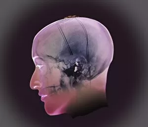
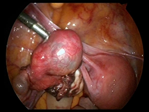
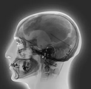
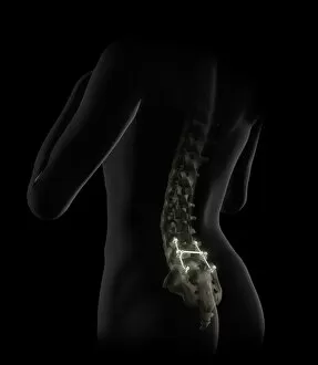
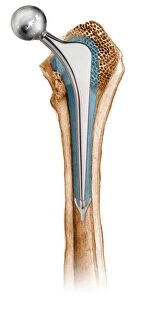
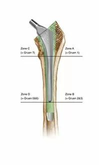
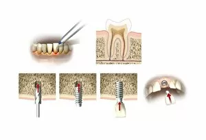
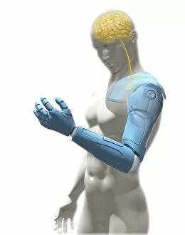
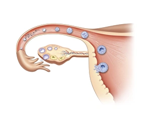
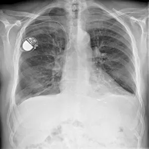
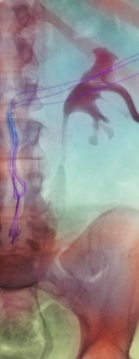
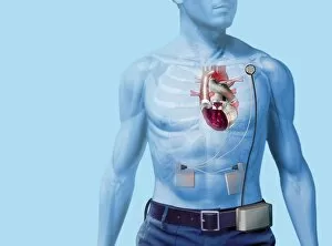
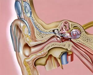
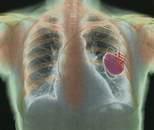
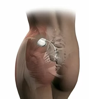
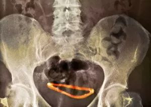
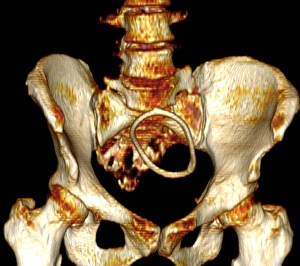
"Implanted: Exploring the Intricacies of Human Intervention" Step into a world where reality blurs with imagination, as we delve into the concept of being "implanted. " Inspired by the legendary Baron Munchausen and his whimsical tales penned by Raspe, this captivating journey takes us through various facets of human existence. Retinal implant artwork captures our attention, reminding us of the incredible advancements in medical technology. It's a testament to how science can restore sight and bring hope to those who once lived in darkness. Intriguingly, an enigmatic portrait writing by Erasmus Rotterdam in 1523 showcases mixed media techniques that hint at hidden messages waiting to be deciphered. Could it be a metaphor for the layers within ourselves that are yet to be explored? The mention of contraceptive implants brings forth thoughts on reproductive choices and personal autonomy. X-ray C017 / 7390 reveals these tiny devices nestled discreetly inside one's body – silent protectors against unwanted pregnancies. Artwork depicting deep brain stimulation captivates our imagination as we ponder upon its potential impact on mental health and neurological disorders. The fusion between artistry and science prompts us to question how far we can push boundaries when it comes to enhancing our cognitive abilities. Cavus foot surgery with metatarsal pinning reminds us that sometimes even mundane procedures can have life-changing effects. This surgical intervention offers individuals suffering from foot deformities newfound freedom and mobility. Ectopic pregnancy viewed through an endoscope (C017 / 6804) highlights both the marvels and challenges faced in modern medicine. It serves as a reminder that despite all progress, there are still mysteries waiting to be unraveled within our own bodies. Ventricular shunt X-rays appear repeatedly, emphasizing their critical role in managing hydrocephalus – a condition affecting fluid balance within the brain. These images remind us of humanity's resilience in finding solutions to complex medical problems.