Fraud Blocker Skip to main content

Invertebrates Collection (page 10)

"Invertebrates

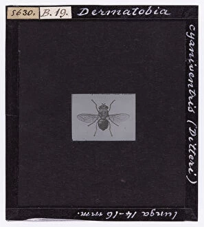
Background imageInvertebrates Collection: Dermatobia cyaniventris (Diptera) from A. Celli, 'Manuale dell'igienista

Dermatobia cyaniventris (Diptera) from A. Celli, "Manuale dell'igienista, ad uso di ufficiali sanitari, medici circondariali e provinciali, ingegneri, chimici e veterinari igienisti

Background imageInvertebrates Collection: Nymph of Culex pipiens (Culicidae), enlarged under the microscope

Nymph of Culex pipiens (Culicidae), enlarged under the microscope. Date of Photograph:04/1914

Background imageInvertebrates Collection: Proboscis fly Eristalis tenax (Syrphidae). Diptera, magnified under the microscope

Proboscis fly Eristalis tenax (Syrphidae). Diptera, magnified under the microscope. Date of Photograph:14/04/1894

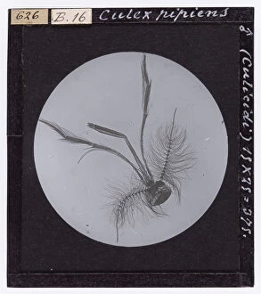
Background imageInvertebrates Collection: Culex pipiens (Culicida. Diptera), magnified under the microscope

Culex pipiens (Culicida. Diptera), magnified under the microscope. Date of Photograph:1914

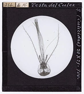
Background imageInvertebrates Collection: Head of female Culex (Diptera) enlarged under a microscope

Head of female Culex (Diptera) enlarged under a microscope. Date of Photograph:1872-1915 ca

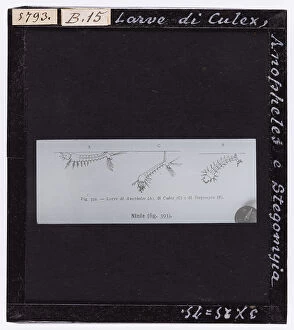
Background imageInvertebrates Collection: Culex, Anopheles and Stegomyia larvae, fig. 392 from A. Celli, Manuale dell'igienista

Culex, Anopheles and Stegomyia larvae, fig. 392 from A. Celli, Manuale dell'igienista, ad uso di ufficiali sanitari, medici circondariali e provinciali, ingegneri, chimici e veterinari igienisti

Background imageInvertebrates Collection: Pulex canis male (Aphaniptera) magnified under the microscope

Pulex canis male (Aphaniptera) magnified under the microscope. Date of Photograph:15/04/1894

Background imageInvertebrates Collection: Sting of the Apis mellifica (Apidae) magnified under the microscope

Sting of the Apis mellifica (Apidae) magnified under the microscope. Date of Photograph:04/1895

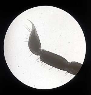
Background imageInvertebrates Collection: Scorpion stinger (Artropodi Scopionoidea) enlarged under a microscope

Scorpion stinger (Artropodi Scopionoidea) enlarged under a microscope Tuscany. Date of Photograph:1878-1898

Background imageInvertebrates Collection: Sting of the Scorpion (Arthropod Scorpionoidea) magnified under the microscope

Sting of the Scorpion (Arthropod Scorpionoidea) magnified under the microscope. Date of Photograph:04/1914

Background imageInvertebrates Collection: Larva of the Crangon Vulgaris (Caridae. Decapodi)enlarged under a microscope

Larva of the Crangon Vulgaris (Caridae. Decapodi)enlarged under a microscope. Date of Photograph:1914

Background imageInvertebrates Collection: Daphnia Magna (Crostacei. Entomostracee) enlarged under a microscope

Daphnia Magna (Crostacei. Entomostracee) enlarged under a microscope. Date of Photograph:19/04/1894

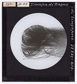
Background imageInvertebrates Collection: Singapore spider leg enlarged under a microscope

Singapore spider leg enlarged under a microscope. Date of Photograph:19/04/1894

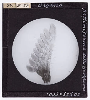
Background imageInvertebrates Collection: Scorpion pectiniform organ magnified under a microscope

Scorpion pectiniform organ magnified under a microscope. Date of Photograph:1888 ca

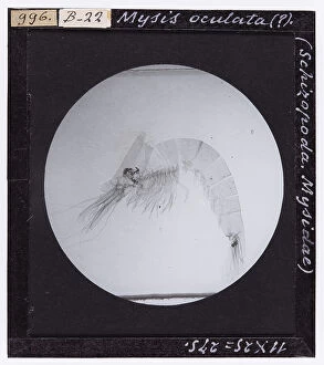
Background imageInvertebrates Collection: Mysis oculata (Schizopoda. Mysidae) magnified under the microscope

Mysis oculata (Schizopoda. Mysidae) magnified under the microscope. Date of Photograph:04/1914

Background imageInvertebrates Collection: Balanus balanoides (Cirripedes, Balanidae) magnified under the microscope

Balanus balanoides (Cirripedes, Balanidae) magnified under the microscope. Date of Photograph:04/1914

Background imageInvertebrates Collection: Mysis spinulosus (Schizopoda, Mysidae) magnified under the microscope

Mysis spinulosus (Schizopoda, Mysidae) magnified under the microscope. Date of Photograph:04/1914

Background imageInvertebrates Collection: Tetranychus persicae (Trombidiidae. Acarina) magnified under the microscope

Tetranychus persicae (Trombidiidae. Acarina) magnified under the microscope. Date of Photograph:1914

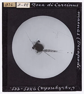
Background imageInvertebrates Collection: Zoea of Carcinus maenas (Decapoda Oxyrhyncha) magnified under the microscope

Zoea of Carcinus maenas (Decapoda Oxyrhyncha) magnified under the microscope. Date of Photograph:05/1895

Background imageInvertebrates Collection: Aphis Rosae (Aphidae-Emipteri) enlarged under a microscope

Aphis Rosae (Aphidae-Emipteri) enlarged under a microscope. Date of Photograph:1914

Background imageInvertebrates Collection: Chrysops coecutiens (Biptera. Brachycera. Tabanidae), from A. Celli, 'Manuale dell'igienista

Chrysops coecutiens (Biptera. Brachycera. Tabanidae), from A. Celli, "Manuale dell'igienista, ad uso di ufficiali sanitari, medici circondariali e provinciali, ingegneri

Background imageInvertebrates Collection: Piophila casei female with larva and nymph (Diptera) 4 mm long, from A

Piophila casei female with larva and nymph (Diptera) 4 mm long, from A. Celli, "Manuale dell'igienista, ad uso di ufficiali sanitari, medici circondariali e provinciali, ingegneri

Background imageInvertebrates Collection: Hypoderma Bovis (Diptera) from A. Celli, 'Manuale dell'igienista, ad uso di ufficiali sanitari

Hypoderma Bovis (Diptera) from A. Celli, "Manuale dell'igienista, ad uso di ufficiali sanitari, medici circondariali e provinciali, ingegneri, chimici e veterinari igienisti

Background imageInvertebrates Collection: Musca domestica and larva, fig. 399 from A. Celli, 'Manuale dell'igienista

Musca domestica and larva, fig. 399 from A. Celli, "Manuale dell'igienista, ad uso di ufficiali sanitari, medici circondariali e provinciali, ingegneri, chimici e veterinari igienisti

Background imageInvertebrates Collection: Glossina palpalis (Diptera. Brachycera. Muscidae), from A. Celli, 'Manuale dell'igienista

Glossina palpalis (Diptera. Brachycera. Muscidae), from A. Celli, "Manuale dell'igienista, ad uso di ufficiali sanitari, medici circondariali e provinciali, ingegneri

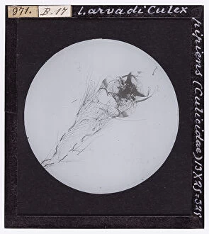
Background imageInvertebrates Collection: Culex pipiens larva (Culicidae), enlarged under the microscope

Culex pipiens larva (Culicidae), enlarged under the microscope. Date of Photograph:04/1914

Background imageInvertebrates Collection: Tracheae of the proboscis of Musca domestica (Muscidae), enlarged under the microscope

Tracheae of the proboscis of Musca domestica (Muscidae), enlarged under the microscope. Date of Photograph:04/1894

Background imageInvertebrates Collection: Male Chironomus plumosus (Culicidiformes), magnified under the microscope

Male Chironomus plumosus (Culicidiformes), magnified under the microscope. Date of Photograph:06/12/1890

Background imageInvertebrates Collection: Head of male and female Culex pipiens and male and female Anopheles claviger

Head of male and female Culex pipiens and male and female Anopheles claviger, magnified under the microscope. Date of Photograph:1890-1914 ca

Background imageInvertebrates Collection: Male Culex pipiens (Culicidae), magnified under the microscope

Male Culex pipiens (Culicidae), magnified under the microscope. Date of Photograph:04/1895

Background imageInvertebrates Collection: Head of female Culex (Culicidae), enlarged under the microscope

Head of female Culex (Culicidae), enlarged under the microscope. Date of Photograph:06/12/1890

Background imageInvertebrates Collection: Head of male Anapheles (Diptera) enlarged under a microscope

Head of male Anapheles (Diptera) enlarged under a microscope. Date of Photograph:1872-1915 ca

Background imageInvertebrates Collection: Larva of Anopheles (Diptera) magnified under the microscope

Larva of Anopheles (Diptera) magnified under the microscope. Date of Photograph:1872-1915 ca

Background imageInvertebrates Collection: Stegomyia fasciata male and female. Yellow fever, fig. 398 from A. Celli, Manuale dell'igienista

Stegomyia fasciata male and female. Yellow fever, fig. 398 from A. Celli, Manuale dell'igienista, ad uso di ufficiali sanitari, medici circondariali e provinciali, ingegneri

Background imageInvertebrates Collection: Phthirius pubis (Pubic Lice) enlarged under a microscope

Phthirius pubis (Pubic Lice) enlarged under a microscope. Date of Photograph:04/1914

Background imageInvertebrates Collection: Microscopically enlarged sting of Vespa vulgaris (Vespidae)

Microscopically enlarged sting of Vespa vulgaris (Vespidae). Date of Photograph:04/1895

Background imageInvertebrates Collection: Musca domestica (Diptera. Muscidae), from A. Celli, 'Manuale dell'igienista

Musca domestica (Diptera. Muscidae), from A. Celli, "Manuale dell'igienista, ad uso di ufficiali sanitari, medici circondariali e provinciali, ingegneri, chimici e veterinari igienisti

Background imageInvertebrates Collection: Male Anopheles maculiformis, magnified under the microscope

Male Anopheles maculiformis, magnified under the microscope. Date of Photograph:1890-1914 ca

Background imageInvertebrates Collection: Pupa of Culex Pipiens magnified under a microscope

Pupa of Culex Pipiens magnified under a microscope. Date of Photograph:04/1914

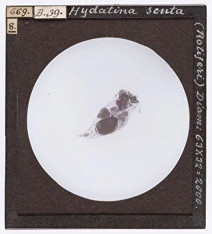
Background imageInvertebrates Collection: Hydatina scrita: microscopic fresh-water animal, belonging to the rotifers

Hydatina scrita: microscopic fresh-water animal, belonging to the rotifers, endowed with two rows of a vibratile mantle situated around the mouth, which are also used for movementt

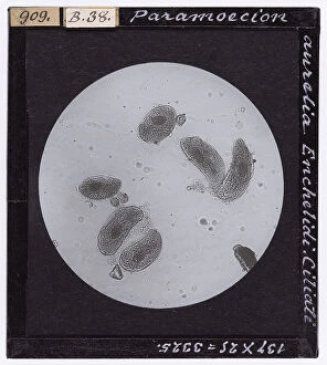
Background imageInvertebrates Collection: Ciliate protozoa, Paramecium aurelia, enlarged under a microscope (Enchelida)

Ciliate protozoa, Paramecium aurelia, enlarged under a microscope (Enchelida). Date of Photograph:1914

Background imageInvertebrates Collection: Ematic alterations in oxen, enlarged under a microscope

Ematic alterations in oxen, enlarged under a microscope. Date of Photograph:1872-1915 ca

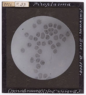
Background imageInvertebrates Collection: Tropical babesia in oxen (Haemosproidia), enlarged under a microscope

Tropical babesia in oxen (Haemosproidia), enlarged under a microscope. Date of Photograph:1872-1915 ca

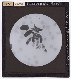
Background imageInvertebrates Collection: Sarcocystis bovis (Sarcosporidia) enlarged under a microscope

Sarcocystis bovis (Sarcosporidia) enlarged under a microscope. Date of Photograph:1872-1915 ca

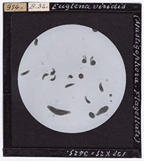
Background imageInvertebrates Collection: Euglena viridis (Mastigophora-Flagellati) enlarged under a microscope

Euglena viridis (Mastigophora-Flagellati) enlarged under a microscope. Date of Photograph:1914

Background imageInvertebrates Collection: Hematococcus nivalis, belonging to the Phitomonadine family, enlarged under a microscope

Hematococcus nivalis, belonging to the Phitomonadine family, enlarged under a microscope. Date of Photograph:04/1913

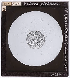
Background imageInvertebrates Collection: Volvox globator: infusorians, flagellati enlarged under a microscope

Volvox globator: infusorians, flagellati enlarged under a microscope. Date of Photograph:1897

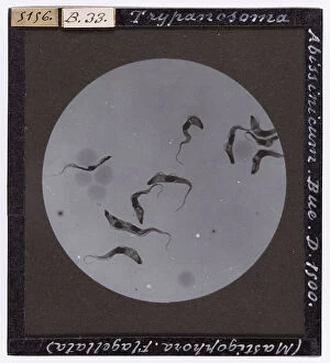
Background imageInvertebrates Collection: Types of protozoa flagellata: Tripanosoma Abissinicum, belonging to the Bovis species

Types of protozoa flagellata: Tripanosoma Abissinicum, belonging to the Bovis species, enlarged under a microscope. Date of Photograph:1872-1915 ca


All Professionally Made to Order for Quick Shipping

"Invertebrates: A Fascinating World of Diversity and Adaptation" From the intricate honeycombs built by diligent honey bees to the captivating sight of a lobster's inner workings revealed through an X-ray, invertebrates never cease to amaze us. The Red Admiral butterfly gracefully displays its vibrant wings, showcasing nature's artistic brilliance. Meanwhile, under the lens of a scanning electron microscope, calcareous phytoplankton reveal their delicate beauty as they float effortlessly in the vast ocean. In Kagoshima Prefecture, Kyushu, Japan, a male Leach's sea star steals the spotlight as it engages in broadcast spawning - an awe-inspiring phenomenon where streams of sperm are released from its arms into the water. This mesmerizing moment captured by a talented wildlife photographer earned them recognition as the winner in the Underwater category at Wildlife Photographer of the Year 2022. Venturing into Croatian waters along Korcula Island in the Adriatic Sea reveals another enchanting creature - a Nudibranch known as Janolus cristatus. Its vibrant colors and graceful movements make it a true gem beneath the waves. A sea green swallowtail butterfly flutters elegantly across meadows and gardens while fruit flies unveil their intricate structures when observed under high-resolution SEM imaging techniques. Delving deep into Earth's history brings us face-to-face with ancient wonders like trilobite fossils that offer glimpses into prehistoric times. These extinct arthropods remind us of our planet's rich evolutionary past. Witnessing an ocellate octopus swimming up from the depths is like witnessing magic unfold before our eyes. Its ethereal appearance leaves us captivated by nature's ability to create such extraordinary creatures. Lastly, we encounter a painted lady butterfly whose delicate patterns adorn fields with splashes of color and gracefulness. And even within microscopic realms lies astonishing complexity; for instance, observing dog tapeworm heads through SEM reveals the intricate structures that allow these parasites to thrive.