Fraud Blocker Skip to main content

Joint Collection (page 9)

"Exploring the intricate world of joints: from normal knees to X-ray revelations

Background imageJoint Collection: Hand anatomy engraving 1866

Hand anatomy engraving 1866
Atlas d anatomie descriptive du corps humain C. Bonamy - Paul Broca Victor Masson et Fils Paris 1866

Background imageJoint Collection: Knee region anatomy engraving 1866

Knee region anatomy engraving 1866
Atlas d anatomie descriptive du corps humain C. Bonamy - Paul Broca Victor Masson et Fils Paris 1866

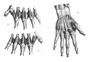
Background imageJoint Collection: Dorsal region of hand engraving 1866

Dorsal region of hand engraving 1866
Atlas d anatomie descriptive du corps humain C. Bonamy - Paul Broca Victor Masson et Fils Paris 1866

Background imageJoint Collection: anatomy engraving 1866

anatomy engraving 1866
Atlas d anatomie descriptive du corps humain C. Bonamy - Paul Broca Victor Masson et Fils Paris 1866

Background imageJoint Collection: Region anterior of arm engraving 1866

Region anterior of arm engraving 1866
Atlas d anatomie descriptive du corps humain C. Bonamy - Paul Broca Victor Masson et Fils Paris 1866

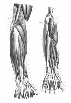
Background imageJoint Collection: Posterior part of forearm engraving 1866

Posterior part of forearm engraving 1866
Atlas d anatomie descriptive du corps humain C. Bonamy - Paul Broca Victor Masson et Fils Paris 1866

Background imageJoint Collection: Region anterior of forearm engraving 1866

Region anterior of forearm engraving 1866
Atlas d anatomie descriptive du corps humain C. Bonamy - Paul Broca Victor Masson et Fils Paris 1866

Background imageJoint Collection: Region external of arm engraving 1866

Region external of arm engraving 1866
Atlas d anatomie descriptive du corps humain C. Bonamy - Paul Broca Victor Masson et Fils Paris 1866

Background imageJoint Collection: Pelvic femoral region anatomy engraving 1866

Pelvic femoral region anatomy engraving 1866
Atlas d anatomie descriptive du corps humain C. Bonamy - Paul Broca Victor Masson et Fils Paris 1866

Background imageJoint Collection: Lumbar region anatomy engraving 1866

Lumbar region anatomy engraving 1866
Atlas d anatomie descriptive du corps humain C. Bonamy - Paul Broca Victor Masson et Fils Paris 1866

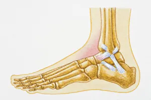
Background imageJoint Collection: Illustration showing swollen talocrural joint caused by sprain to anterior talofibular ligament

Illustration showing swollen talocrural joint caused by sprain to anterior talofibular ligament

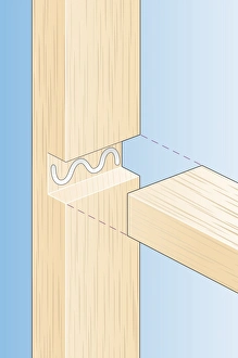
Background imageJoint Collection: Digital Illustration of bridle joint in wood

Digital Illustration of bridle joint in wood

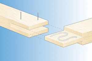
Background imageJoint Collection: Digital Illustration of adhesive inside join of housing joint in wood

Digital Illustration of adhesive inside join of housing joint in wood

Background imageJoint Collection: Digital illustration of repointing mortar joint around metal flashing above extension roof using

Digital illustration of repointing mortar joint around metal flashing above extension roof using trowel

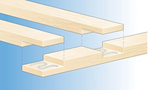
Background imageJoint Collection: Digital illustration of gluing wood halving joints to end and middle of timber

Digital illustration of gluing wood halving joints to end and middle of timber

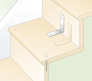
Background imageJoint Collection: Digital illustration of reinforcing joints between riser and tread using covered screws

Digital illustration of reinforcing joints between riser and tread using covered screws and recessed brackets

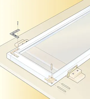
Background imageJoint Collection: Digital illustration of securing loose joints in casement window with recessed metal angle

Digital illustration of securing loose joints in casement window with recessed metal angle and glued dowels driven into mortise-and-tenon joints

Background imageJoint Collection: Illustration of rebate or joint in wood

Illustration of rebate or joint in wood

Background imageJoint Collection: CABIN CONSTRUCTION, 1935. A detail of the construction of a cabin in Corbin Hollow

CABIN CONSTRUCTION, 1935. A detail of the construction of a cabin in Corbin Hollow, Shenandoah National Park, Virginia. Photograph by Arthur Rothstein, October 1935

Background imageJoint Collection: NOVA BRITANNIA, 1609. Title-page of Nova Britannia, the pamphlet issued in 1609

NOVA BRITANNIA, 1609. Title-page of Nova Britannia, the pamphlet issued in 1609 by the Virginia Company in London to win support for their enterprise in America

Background imageJoint Collection: Conceptual image of human legs and feet with nervous system

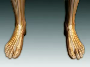
Conceptual image of human legs and feet with nervous system

Background imageJoint Collection: Detail of human knee showing insertion of arthroscopic instruments

Detail of human knee showing insertion of arthroscopic instruments

Background imageJoint Collection: Sciatica caused from herniated disc

Sciatica caused from herniated disc

Background imageJoint Collection: X-ray view of human foot

X-ray view of human foot

Background imageJoint Collection: Longitudinal section of human foot in a sagittal plane

Longitudinal section of human foot in a sagittal plane

Background imageJoint Collection: Illustration of knee bone, bending

Illustration of knee bone, bending

Background imageJoint Collection: Conceptual image of pain in human foot

Conceptual image of pain in human foot

Background imageJoint Collection: Anatomy of acromioclavicular joint rupture and displacement

Anatomy of acromioclavicular joint rupture and displacement

Background imageJoint Collection: X-ray image of knee

X-ray image of knee

Background imageJoint Collection: Bones in the human foot

Bones in the human foot

Background imageJoint Collection: Conceptual image of gout in the big toe

Conceptual image of gout in the big toe. Gout is a form of inflammatory arthritis that causes sudden, severe pain, swelling and tenderness of joints

Background imageJoint Collection: Anatomy of the dog elbow with lateral zoom

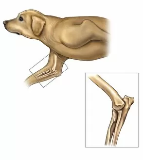
Anatomy of the dog elbow with lateral zoom

Background imageJoint Collection: Anatomy of human pelvic bone and ligaments

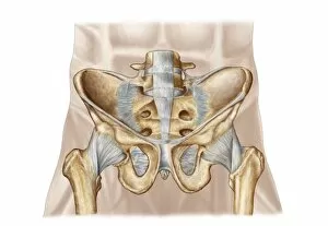
Anatomy of human pelvic bone and ligaments

Background imageJoint Collection: Head of femur fracture

Head of femur fracture

Background imageJoint Collection: Illustration of knee bone, straight

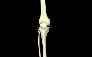
Illustration of knee bone, straight

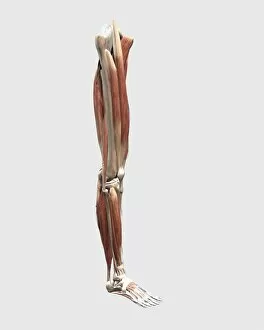
Background imageJoint Collection: Medical illustration of human leg muscles, bones and joints

Medical illustration of human leg muscles, bones and joints

Background imageJoint Collection: Conceptual image of muscle cramp and pain in human foot

Conceptual image of muscle cramp and pain in human foot

Background imageJoint Collection: Dorsal anatomy of the human foot

Dorsal anatomy of the human foot

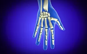
Background imageJoint Collection: Conceptual image of bones in human hand

Conceptual image of bones in human hand

Background imageJoint Collection: Typical synovial joint

Typical synovial joint

Background imageJoint Collection: Detail of human knee showing arthroscopic surgical proceedures

Detail of human knee showing arthroscopic surgical proceedures

Background imageJoint Collection: Anterior compartment anatomy of left leg muscles and tendons

Anterior compartment anatomy of left leg muscles and tendons

Background imageJoint Collection: Anatomy of left shoulder, coronal view

Anatomy of left shoulder, coronal view


All Professionally Made to Order for Quick Shipping

"Exploring the intricate world of joints: from normal knees to X-ray revelations. " Witness the marvels of our body's architecture as we delve into the anatomy of a human knee joint, where strength and flexibility meet. Step into Shoreham-by-Sea's bustling High Street and discover a hidden gem – the Butchers Shop, where skilled hands expertly carve beef joints for your culinary delight. Unveiling Marie Curie's groundbreaking discoveries in science, she proved that knowledge is indeed a powerful joint between passion and dedication. The Vegetabull proudly presents "The Joint Poster, " showcasing an array of vibrant vegetables coming together harmoniously to create delicious meals bursting with flavor. Market Reports reveal how English Country Squires skillfully carve beef joints, ensuring every slice is perfection on a plate fit for royalty. Hip replacement becomes art as talented artists capture its essence through stunning artwork, reminding us that even in adversity, beauty can emerge. Peering through an X-ray lens reveals the wonders of modern medicine - witness the transformation from pain to mobility with a hip joint replacement. Delve into nature's microcosm as we explore an ant under scanning electron microscopy (SEM), highlighting their remarkable ability to navigate their tiny yet complex joints effortlessly. A closer look at bunions through X-ray imagery reminds us that even small imperfections can impact our daily lives; seek proper care and relief when needed. Shoulder tendinitis captured by X-ray serves as a reminder to nurture our bodies while pushing boundaries – listen closely when they whisper about overuse or strain.