
Keratinocyte Collection
Keratinocytes, the main cells found in the epidermis, play a crucial role in maintaining healthy skin


All Professionally Made to Order for Quick Shipping
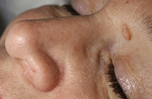
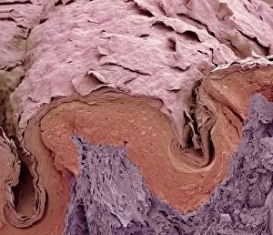
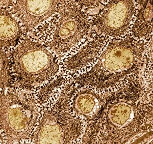
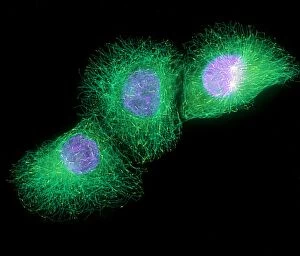
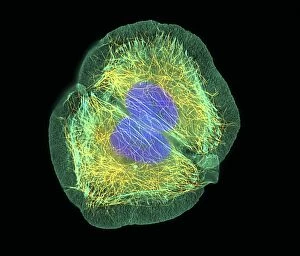
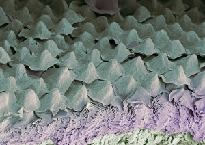
Keratinocytes, the main cells found in the epidermis, play a crucial role in maintaining healthy skin. These specialized skin cells are responsible for producing keratin, a tough protein that forms the protective barrier of our skin. Under the powerful lens of a transmission electron microscope (TEM), we can observe these keratinocytes up close and personal. Their intricate structure is revealed, showcasing their importance in safeguarding our body from external threats. Dermal nevi, commonly known as moles or birthmarks, often appear raised on the surface of the skin. When examined using scanning electron microscopy (SEM), these elevated patches become more pronounced and detailed. The SEM images provide us with an insight into their unique characteristics and how they differ from surrounding skin. Moving to another part of our body - fingers. By utilizing SEM once again, we can delve into the fascinating world of finger skin at an incredibly high resolution. The intricate patterns and textures become evident under this microscopic view, highlighting just how complex even seemingly simple things like fingerprints truly are. Zooming further into focus through SEM imagery specifically labeled C013 / 4781 and C013 / 4780, we witness individual keratinocyte skin cells magnified to astonishing detail. Each cell appears like a tiny fortress protecting our inner layers from harm – truly remarkable. Not limited to just one type of cell though; TEM allows us to explore various types of skin cells present within our bodies. From general views showing multiple interconnected cells to highly detailed images focusing on single entities – all revealing different aspects essential for understanding overall tissue health. When injuries occur on our delicate dermis layer resulting in wound scabs forming over time; SEM provides us with valuable insights into their composition and structure as well. These captivating images showcase how keratinocytes work together during healing processes by forming protective barriers until complete restoration is achieved. Exploring keratinocyte-rich tissues through advanced imaging techniques such as TEM and SEM allows us to appreciate the intricate beauty of our skin cells.