
Labeled Collection (page 3)
"Exploring the World of Labels: From Art to Science" Delve into the intricate rhythms of life with ECGs showcasing a normal heart rate, beautifully captured in artwork


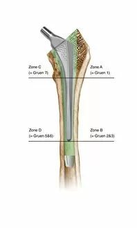


All Professionally Made to Order for Quick Shipping
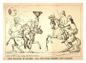
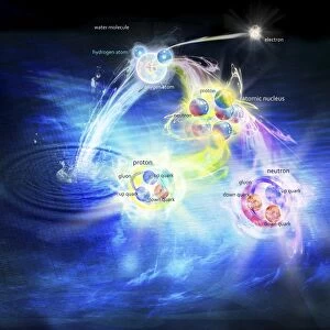
"Exploring the World of Labels: From Art to Science" Delve into the intricate rhythms of life with ECGs showcasing a normal heart rate, beautifully captured in artwork. Uncover the wonders of our vision as we unravel the mysteries behind eye anatomy through stunning artistic representations. Embark on an awe-inspiring journey through time and space with a captivating universe timeline depicted in mesmerizing artwork. Witness history being made as you trace the orbit of Sputnik 1, Soviet's groundbreaking achievement in 1957, illustrated in a detailed diagram. Dive into the microscopic world and marvel at Trypanosome protozoan brought to life through exquisite artwork. Discover the building blocks of our body's strength - collagen synthesis and assembly - portrayed artistically for your visual pleasure. Get up close and personal with fibroblast cells, essential contributors to tissue repair and regeneration, showcased in stunning artwork. Experience the beauty behind muscle contraction as it is elegantly depicted through artistry that captures its power and grace. Find solace amidst thorns with Gillray's iconic engraving "Comfort of a Bed of Roses, " offering a satirical take on life's challenges during his era (1756-1815). Explore human anatomy from every angle as you delve into lower back muscles intricately labeled in an illustrative masterpiece. Peer inside animal cell structures like never before, unlocking their secrets through visually striking depictions that captivate your imagination. Relive history with a poignant photograph capturing "The Situation at the Battle of Wounded Knee" when tensions erupted between Native Americans and U. S forces in 1891. Step into this world where art meets science; let these labeled artworks guide you on an extraordinary voyage filled with knowledge, inspiration, and wonderment.