
Lacunae Collection
"Lacunae: Unveiling the Intricate World of Microscopic Spaces" Delving into the depths of nature's secrets


All Professionally Made to Order for Quick Shipping
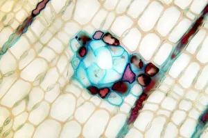
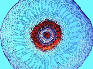
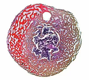
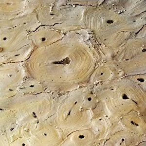
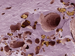
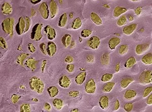
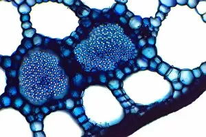
"Lacunae: Unveiling the Intricate World of Microscopic Spaces" Delving into the depths of nature's secrets, a pine stem reveals its hidden beauty under the lens of a light micrograph. Lacunae, small cavities within this structure, hold untold stories waiting to be discovered. Journeying further into the realm of bone, a compact bone unveils its intricate network through a mesmerizing light micrograph. Within these lacunae lies an unseen world where cells reside and communicate with each other. Like delicate water lily leaves floating on calm waters, their enchanting beauty extends beyond what meets the eye. A light micrograph exposes lacunae within these leaves, offering glimpses into their complex cellular architecture. Beneath the surface of a water fern rhizome lies an astonishing labyrinth of interconnected spaces known as lacunae. Through a captivating light micrograph, we witness nature's ingenuity in creating structures that support life. The spongy bone dances with grace under the microscope's gaze, revealing its porous texture and countless lacunae housing living cells responsible for maintaining our skeletal framework. Exploring deeper into aquatic realms brings us to pondweed stems adorned with intricate patterns when viewed through a light micrograph—each pattern representing unique arrangements that contribute to plant growth and survival. Stepping away from microscopic wonders but still embracing artistry is "Episodes from the Life of St. Augustine, " depicted in fresco form between 1463-65 by unknown hands (see also 192535, 192549). Just like hidden lacunae within bones or plants' tissues reveal secrets about their existence; this artwork hides tales waiting to be deciphered by curious minds. Captivating our attention once again is compact bone—a marvelously structured tissue revealed through both light micrographs and scanning electron microscopy (SEM).