Fraud Blocker Skip to main content

Leukocyte Collection (page 8)

"Exploring the Mighty Defenders: Leukocytes and their Battle against Disease" In this captivating collection of images, we delve into the fascinating world of leukocytes

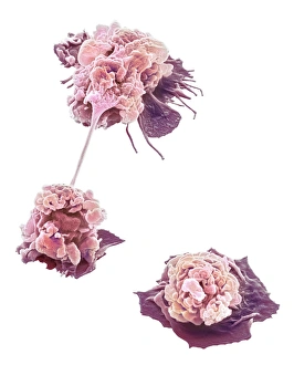
Background imageLeukocyte Collection: Activated granulocytes, SEM

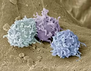
Activated granulocytes, SEM
Activated granulocytes. Coloured scanning electron micrograph (SEM) of activated granulocyte, or polymorphonucleocytes (PMNs), white blood cells

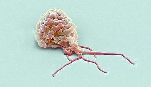
Background imageLeukocyte Collection: Activated granulocyte, SEM

Activated granulocyte, SEM
Activated granulocyte. Coloured scanning electron micrograph (SEM) of an activated granulocyte, or polymorphonucleocyte (PMN), white blood cell

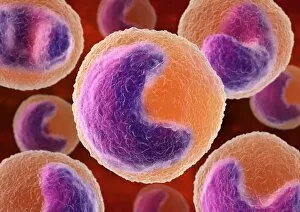
Background imageLeukocyte Collection: Monocyte white blood cells, artwork

Monocyte white blood cells, artwork
Monocyte white blood cells. Computer artwork of monocyte white blood cells, showing their lobed nuclei (purple, centre). Monocytes, like all white blood cells

Background imageLeukocyte Collection: Basophil white blood cells, artwork

Basophil white blood cells, artwork
Basophil white blood cells. Computer artwork of basophil white blood cells, showing their lobed nuclei (red). Basophils are the smallest and least common of the white blood cells

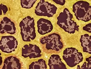
Background imageLeukocyte Collection: Lymph node, TEM

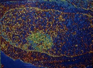
Lymph node, TEM
Lymph node. Coloured transmission electron micrograph (TEM) of a section through the peripheral corona of a lymph nodule, showing numerous small lymphocyte white blood cells (dark)

Background imageLeukocyte Collection: Eosinophil white blood cells, artwork

Eosinophil white blood cells, artwork
Eosinophil white blood cells. Computer artwork of eosinophil white blood cells, showing their lobed nuclei (centre). Eosinophils, like all white blood cells, are part of the immune system

Background imageLeukocyte Collection: Cell infected with HIV, electronic image

Cell infected with HIV, electronic image
Experts of the Ivanovsky Virusology Institute [USSR Academy of Medical Sciences] have made this electronic image of a cell infected with the AIDS virus

Background imageLeukocyte Collection: Macrophage white blood cell, TEM

Macrophage white blood cell, TEM
Macrophage white blood cell. Coloured transmission electron micrograph (TEM) of a section through a macrophage from a lymph node, showing the nucleus (red) and numerous lysosomes (black)

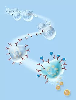
Background imageLeukocyte Collection: Immune system, artwork

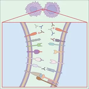
Immune system, artwork
Immune system. Computer artwork showing how T-cells (right), antibodies (Y-shaped) and antigen-presenting cells (APC, left) interact during an immune response

Background imageLeukocyte Collection: Human immune response, artwork

Human immune response, artwork
Human immune response. Computer artwork showing the interactions of the various white blood cells (purple) involved during an immune response in the human body

Background imageLeukocyte Collection: Immune response to chronic inflammation

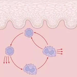
Immune response to chronic inflammation. Computer artwork showing the interaction of T cell, lymphocyte, and macrophage white blood cells in response to chronic inflammation of the skin (top)

Background imageLeukocyte Collection: Skin damage immune response, artwork

Skin damage immune response, artwork
Skin damage immune response. Computer artwork showing the bodys immune response to pathogens (red dots) entering through a skin wound (upper centre)

Background imageLeukocyte Collection: Blood cells, artwork

Blood cells, artwork
Blood cells. Cut-away artwork of a blood vessel showing the blood cells inside. The majority of blood cells are erythrocytes (red blood cells)

Background imageLeukocyte Collection: Granulocytes, SEM

Granulocytes, SEM
Granulocytes. Coloured scanning electron micrograph (SEM) of granulocytes in bone marrow. Granulocytes are a category of white blood cell characterized by the presence of granules in their cytoplasm

Background imageLeukocyte Collection: Woundcare gel therapy, artwork

Woundcare gel therapy, artwork
Woundcare gel therapy. Artwork showing the cellular effects of woundcare therapy using a platelet-leucocyte enriched gel to support soft tissue and bone healing

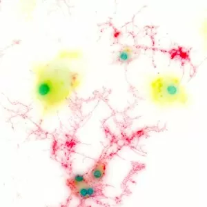
Background imageLeukocyte Collection: Brain cells, light micrograph

Brain cells, light micrograph
Brain cells. Fluorescence light micrograph of activated microglial cells (stained for membrane co-factor protein, yellow) and oligodendrocytes (stained for myelin basic protein, red)

Background imageLeukocyte Collection: Allergic response, artwork

Allergic response, artwork
Allergic response. Computer artwork showing the process that produces an allergic reaction. Antigens (blue), such as pollen

Background imageLeukocyte Collection: Blood cells, SEM

Blood cells, SEM
Blood cells. Coloured scanning electron micrograph (SEM) of a human red blood cell (erythrocyte, red) and a white blood cell (leucocyte, blue). These are the two main cell components of blood

Background imageLeukocyte Collection: Blood cells in blood vessel, artwork

Blood cells in blood vessel, artwork
Blood cells in blood vessel, computer artwork. Red blood cells (erythrocytes, red) distribute oxygen to body tissues, and carry waste carbon dioxide back to the lungs

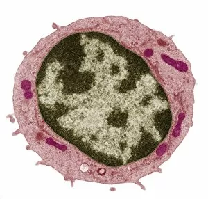
Background imageLeukocyte Collection: Small lymphocyte, TEM

Small lymphocyte, TEM
Small lymphocyte. Coloured transmission electron micrograph (TEM) showing the large central nucleus (brown) of a small lymphocyte (white blood cell)

Background imageLeukocyte Collection: White blood cell, TEM

White blood cell, TEM
White blood cell. Coloured transmission electron micrograph (TEM) of an eosinophil (a type of white blood cell), part of the bodys immune system

Background imageLeukocyte Collection: Dead neutrophil white blood cell, SEM

Dead neutrophil white blood cell, SEM
Dead neutrophil white blood cell. Coloured scanning electron micrograph (SEM) of a dead neutrophil (green) surrounded by rod-shaped bacteria (blue). Neutrophils are part of the bodys immune response

Background imageLeukocyte Collection: Human blood cells, SEM

Human blood cells, SEM
Human blood cells, coloured scanning electron micrograph (SEM). Seen here are platelets (small, roundish) and a neutrophil white blood cell (large, whitish)

Background imageLeukocyte Collection: Blood cells in a kidney, SEM

Blood cells in a kidney, SEM
Blood cells in a kidney. Transmission electron micrograph (TEM) of a section through a kidney glomerulus, showing red blood cells (erythrocytes, red) and a white blood cell (leucocyte, blue)

Background imageLeukocyte Collection: Cell infected with HIV, SEM

Cell infected with HIV, SEM
Cell infected with HIV. Coloured scanning electron micrograph (SEM) of HIV particles (yellow) budding from the membrane of a host cell (red)


All Professionally Made to Order for Quick Shipping

"Exploring the Mighty Defenders: Leukocytes and their Battle against Disease" In this captivating collection of images, we delve into the fascinating world of leukocytes, our body's mighty defenders against various diseases. First up, we witness the incredible power of T lymphocytes as they engage in a fierce battle with cancer cells. SEM C001 / 1679 reveals an epic showdown between these two forces. Moving on to another remarkable image, Neutrophil engulfing MRSA (SEM C018 / 8596) showcases the extraordinary ability of neutrophils to devour harmful bacteria such as Methicillin-resistant Staphylococcus aureus. Next, TEM unveils a close-up view of a human white blood cell bearing HLA antigen. This intricate image highlights the complexity and diversity within our immune system. A burst of color awaits us in Coloured SEM of a white blood cell (lymphocyte), showcasing the vibrant nature that lies within these essential cells. Meanwhile, Basophil white blood cell reminds us that even amidst chaos, there are specialized warriors ready to combat any threat. The delicate balance between red and white blood cells is beautifully captured in Red and white blood cells (SEM). This harmonious coexistence ensures our bodies remain healthy and functional. Micrograph enthusiasts will appreciate Dohle bodies in blood cell micrograph - an intriguing glimpse into abnormal cellular structures that can indicate certain health conditions like infections or inflammation. Shifting gears towards Plasma cells (TEM), we witness their crucial role in producing antibodies to fight off infections effectively. Their unique appearance underlines their significance in maintaining our overall well-being. Acute promyelocytic leukemia takes center stage next with its distinct features showcased through a powerful micrograph. This visual representation sheds light on this specific type of cancer affecting immature myeloid cells. Phagocytosis takes center stage once again as fungal spores become prey for vigilant macrophages in Phagocytosis of fungal spores (SEM).