Fraud Blocker Skip to main content

Light Micrograph Collection

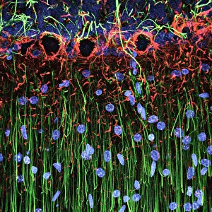
Capturing the intricate beauty of biological structures, a light micrograph reveals the mesmerizing complexity of cerebellum tissue

Background imageLight Micrograph Collection: Cerebellum tissue, light micrograph

Cerebellum tissue, light micrograph
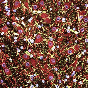
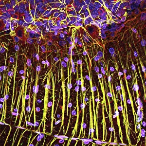
Cerebellum tissue. Confocal light micrograph of a section through the cerebellum of the brain. Purkinje cells, a type of neuron (nerve cell), are red

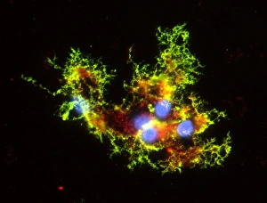
Background imageLight Micrograph Collection: Nerve and glial cells, light micrograph

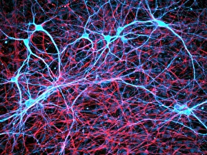
Nerve and glial cells, light micrograph
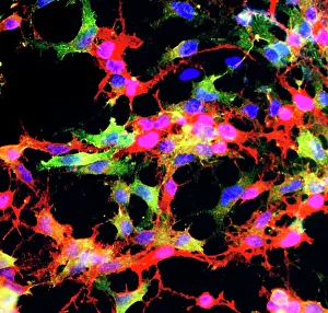
Nerve and glial cells, fluorescence light micrograph. These are neural stem cells that have differentiated into neurons (nerve cells, blue) and glial cells (support cells, red)

Background imageLight Micrograph Collection: Caffeine crystals, light micrograph

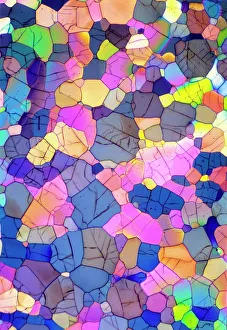
Caffeine crystals, light micrograph
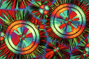
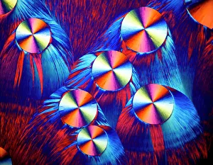
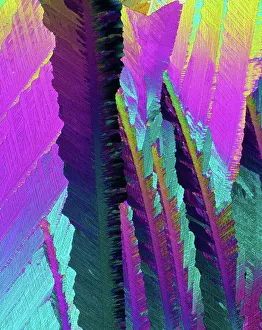
Caffeine crystals. Polarised light micrograph of crystals of caffeine (1, 3, 7-trimethylxanthine). Caffeine stimulates the central nervous system increasing alertness and deferring fatigue

Background imageLight Micrograph Collection: Copper and magnesium sulphate, LM

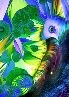
Copper and magnesium sulphate, LM
Copper and magnesium sulphate crystals. Polarised light micrograph (LM) of copper sulphate (CuSO4) and magnesium sulphate (MgSO4) crystals

Background imageLight Micrograph Collection: Hippocampus brain tissue

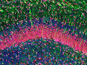
Hippocampus brain tissue
Hippocampus tissue. Light micrograph of a sagittal (side view) section through the hippocampus of the brain showing the nerve cells within it

Background imageLight Micrograph Collection: Immunofluorescent LM of neurons & astrocytes

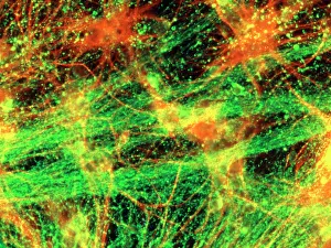
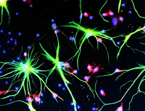
Immunofluorescent LM of neurons & astrocytes
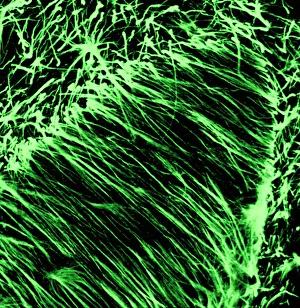
Immunofluorescent Light Micrograph of a network of neurons and astrocyte cells, in brain cortex. In the foreground, nerve fibres of neurons (green) are seen in a fine branching network

Background imageLight Micrograph Collection: HeLa cells, light micrograph C017 / 8299

HeLa cells, light micrograph C017 / 8299
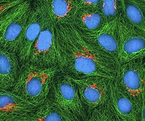
HeLa cells, multiphoton fluorescence micrograph (MFM). The cell nuclei, which contain the cells genetic information, are purple. Microtubules are blue and actin microfilaments are red

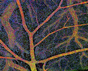
Background imageLight Micrograph Collection: Brain tissue blood supply

Brain tissue blood supply. Light micrograph of a section through cortex tissue from a brain, showing the blood vessels (branching) that supply it

Background imageLight Micrograph Collection: Dicotyledon plant stem, light micrograph

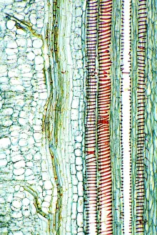
Dicotyledon plant stem, light micrograph
Dicotyledon plant stem. Light micrograph of a longitudinal radial section through a typical dicotyledon stem. The section has passed through a vascular bundle and other stem tissue

Background imageLight Micrograph Collection: EDTA crystals, light micrograph

EDTA crystals, light micrograph
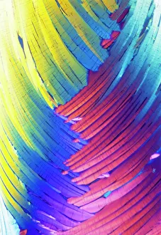
EDTA crystals. Polarised light micrograph of a section through ethylenediaminetetraacetic acid (EDTA) crystals. EDTA is used primarily for its effectiveness at binding metal ions (chelating)

Background imageLight Micrograph Collection: Human blastocyst

Human blastocyst
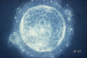
Light micrograph of a human blastocyst, an early stage of embryonic development, photographed 4-5 days after fertilisation

Background imageLight Micrograph Collection: Optical computing

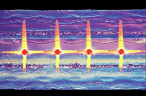
Optical computing. Coloured light micrograph of a section through a hybrid electronic-optical circuit board. Optical waveguides (red) are used to transmit data in the form of light pulses

Background imageLight Micrograph Collection: Polarised LM of a thin section of gabbro rock

Polarised LM of a thin section of gabbro rock
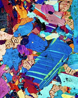
Gabbro. Polarised light micrograph of a thin section of gabbro, an igneous rock. Gabbro is coarse-grained, containing the minerals olivine and plagioclase feldspar

Background imageLight Micrograph Collection: Glial stem cell culture, light micrograph

Glial stem cell culture, light micrograph
Glial stem cell culture. Fluorescent light micrograph of glial stem cells producing the protein NG2 (red) as they mature. These stem cells can differentiate into several types of glial cells

Background imageLight Micrograph Collection: Castor oil stem, light micrograph

Castor oil stem, light micrograph
Castor oil stem. Light micrograph of a longitudinal section through the stem of a castor oil (Ricinus communis) plant. At right are large and small parenchyma cells (blue)

Background imageLight Micrograph Collection: Cerebral cortex nerve cells

Cerebral cortex nerve cells. Confocal light micrograph of neurons (nerve cells, red) and glial cells (support cells, gold) from the cerebral cortex

Background imageLight Micrograph Collection: HeLa cells, light micrograph C017 / 8298

HeLa cells, light micrograph C017 / 8298
HeLa cells, multiphoton fluorescence micrograph (MFM). The cell nuclei, which contain the cells genetic information, are blue. Golgi bodies, which modify and package proteins, are orange

Background imageLight Micrograph Collection: Mitosis, light micrograph

Mitosis, light micrograph
Mitosis. Confocal light micrograph of the stages of mitosis (nuclear division) and cytokinesis (cell division). During mitosis the nuclear envelope disintegrates (3rd image) and the chromosomes (blue)

Background imageLight Micrograph Collection: Lactating breast tissue, light micrograph

Lactating breast tissue, light micrograph
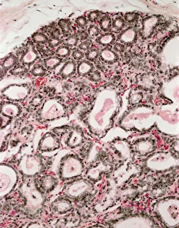
Lactating breast tissue. Light micrograph of a section through healthy breast (mammary gland) tissue during lactation. Glandular tissue in the breast has proliferated to produce spaces (alveoli)

Background imageLight Micrograph Collection: Nerve cells

Nerve cells
Immunofluorescent Light Micrograph of neuron cells and astrocytes in mammalian spinal cord. Here, neuron cells stain red: the cell body appears pink

Background imageLight Micrograph Collection: Microchip, light micrograph

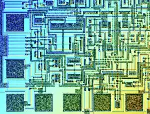
Microchip, light micrograph
Microchip. Light micrograph of the surface of a microchip using differential interference contrast microscopy (DIC). Magnification: x100

Background imageLight Micrograph Collection: Pine pollen grains, light micrograph

Pine pollen grains, light micrograph
Pine pollen grains. Light micrograph of a section through winged pollen grains (microspores) from a pine (Pinus sp.) tree

Background imageLight Micrograph Collection: Pine stem, light micrograph

Pine stem, light micrograph
Pine stem. Light micrograph of a section through the stem of a pine (Pinus sp.) tree, showing xylem tissue. The xylem is made up of tracheid cells (light pink)

Background imageLight Micrograph Collection: Lime tree stem, light micrograph

Lime tree stem, light micrograph
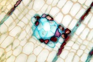
Lime tree stem. Light micrograph of a section through the stem of a lime tree (Tilia europaea). The outer epidermis has been shed and replaced by a layer of cork (dark red)

Background imageLight Micrograph Collection: C. elegans worms, light micrograph

C. elegans worms, light micrograph
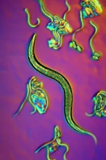
C. elegans worms. Differential interference contrast micrograph of an adult Caenorhabditis elegans worm (centre) surrounded by C. elegans larvae hatching from eggs

Background imageLight Micrograph Collection: Cell division, fluorescent micrograph

Cell division, fluorescent micrograph
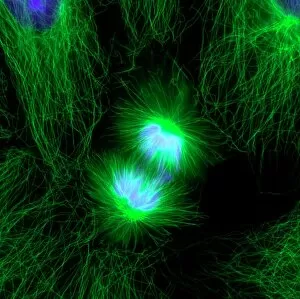
Cell division. Immunofluorescent light micrograph of a human epithelial cell (centre) during the late anaphase stage of mitosis

Background imageLight Micrograph Collection: Neural stem cell culture

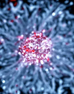
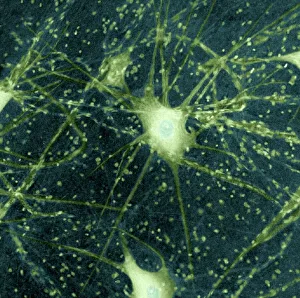
Neural stem cell culture. Fluorescent light micrograph of a group of neural stem cells (neurosphere) in culture. Neural stem cells are able to differentiate into neurons (nerve cells)

Background imageLight Micrograph Collection: Glial cells, confocal light micrograph

Glial cells, confocal light micrograph
Glial cells. Confocal light micrograph of glial cells from the cerebellum of the brain. Glial cells are nervous system cells that provide structural support and protection for neurons (nerve cells)

Background imageLight Micrograph Collection: Compact bone, light micrograph

Compact bone, light micrograph
Compact bone. Polarised light micrograph of a transverse section through compact bone tissue, showing Haversian canals (circular regions)

Background imageLight Micrograph Collection: Copper sulphate crystals, LM

Copper sulphate crystals, LM
Copper sulphate crystals. Polarised light micrograph (LM) of copper sulphate (CuSO4) crystals

Background imageLight Micrograph Collection: Cortisol crystals, light micrograph

Cortisol crystals, light micrograph
Cortisol crystals, polarised light micrograph. Cortisol is a steroid hormone produced by the adrenal glands, which sit on top of the kidneys

Background imageLight Micrograph Collection: Motor neurons, light micrograph

Motor neurons, light micrograph. Motor neurons are responsible for passing information around the central nervous system (CNS) and from the CNS to the rest of the body

Background imageLight Micrograph Collection: Cerebellum tissue, light micrograph

Cerebellum tissue, light micrograph
Cerebellum tissue. Confocal light micrograph of a section through the cerebellum of the brain showing two types of glial cells (support cells); astrocytes (star-shaped)

Background imageLight Micrograph Collection: Dohle bodies in blood cell, micrograph

Dohle bodies in blood cell, micrograph
Dohle bodies in blood cell. Light micrograph of a neutrophil white blood cell (centre) with Dohle bodies. These are small inclusions within the cells cytoplasm
Background imageLight Micrograph Collection: Water bear, light micrograph C016 / 8581

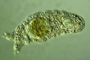
Water bear, light micrograph C016 / 8581
Water bear, differential interference contrast (DIC) light micrograph. Water bears, or tardigrades, (phylum Tardigrada) are small, water-dwelling

Background imageLight Micrograph Collection: Caffeine crystals, light micrograph

Caffeine crystals, light micrograph
Caffeine crystals. Polarised light micrograph of crystals of caffeine (1, 3, 7- trimethylxanthine). Caffeine stimulates the central nervous system increasing alertness and deferring fatigue

Background imageLight Micrograph Collection: Maize root, light micrograph

Maize root, light micrograph
Maize root. Light micrograph of a section through the root of a maize plant (Zea mays) showing a vascular cylinder (centre)

Background imageLight Micrograph Collection: Water lily leaf, light micrograph

Water lily leaf, light micrograph
Water lily leaf. Light micrograph of a transverse section through the leaf of a water lily (Nympha sp.) plant. All aquatic plants (hydrophytes) have a similar structure

Background imageLight Micrograph Collection: Cerebellum tissue, light micrograph

Cerebellum tissue, light micrograph
Cerebellum tissue. Confocal light micrograph of a section through the cerebellum of the brain. Purkinje cells, a type of neuron (nerve cell), are red

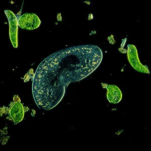
Background imageLight Micrograph Collection: Light Micrograph (LM): Protozoans: Kidney shaped ciliate surrounded by Euglena sp

Light Micrograph (LM): Protozoans: Kidney shaped ciliate surrounded by Euglena sp
LRDS-316 Light Micrograph (LM): Protozoans: Kidney shaped ciliate surrounded by Euglena sp. Magnification x 900 (when printed A4, 29.7 cm wide) Tillina sp

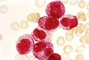
Background imageLight Micrograph Collection: Acute promyelocytic leukaemia, micrograph

Acute promyelocytic leukaemia, micrograph
Acute promyelocytic leukaemia. Light micrograph of blood cells from bone marrow in a case of acute promyelocytic leukaemia. Leukaemia is a cancer where certain blood cells form in excess

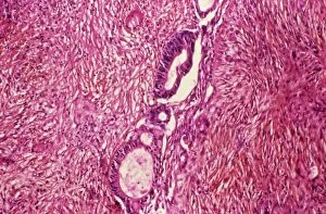
Background imageLight Micrograph Collection: Ovarian cancer, light micrograph C015 / 7103

Ovarian cancer, light micrograph C015 / 7103
Ovary cancer. Light micrograph of a section through a Krukenberg tumour (down centre) of the ovary. This is a secondary cancer that spreads (metastasises) from the digestive system

Background imageLight Micrograph Collection: Seed shrimp, light micrograph

Seed shrimp, light micrograph
Seed shrimp. Light micrograph of a seed shrimp (Cyclocypris sp.). The body of this freshwater crustacean is enclosed in two shells, which it can open and close using a special muscle

Background imageLight Micrograph Collection: Polarised LM of a molar tooth showing decay

Polarised LM of a molar tooth showing decay
Tooth decay. Polarised light micrograph (PLM) of a longitudinal section through a molar tooth with dental caries, or tooth decay

Background imageLight Micrograph Collection: Oligodendrocyte nerve cells

Oligodendrocyte nerve cells. Fluorescent light micrograph of human oligodendrocyte nerve cells. Cell nuclei, which contain the cells genetic information, have been dyed blue

Background imageLight Micrograph Collection: Liver tissue cirrhosis, light micrograph

Liver tissue cirrhosis, light micrograph
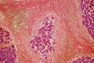
Liver cirrhosis. Light micrograph of a section through liver tissue showing cirrhosis. This is a condition where the liver responds to the injury or death of some of its cells by producing

Background imageLight Micrograph Collection: Cystic fibrosis

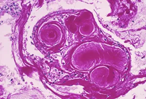
Cystic fibrosis. Light micrograph of a section through a bronchus (airway) of the lungs in a case of cystic fibrosis (mucoviscidosis)

Background imageLight Micrograph Collection: Prostate, light micrograph

Prostate, light micrograph
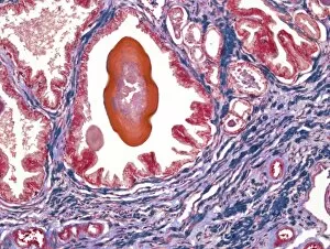
Prostate. Light micrograph of a section through a prostate showing a concretion (red) of secretory materials in a prostatic alveolus (air space)


All Professionally Made to Order for Quick Shipping

Capturing the intricate beauty of biological structures, a light micrograph reveals the mesmerizing complexity of cerebellum tissue. Delicate nerve and glial cells intertwine like a finely woven tapestry, forming the foundation for neurological function. In another stunning image, copper and magnesium sulphate crystals shimmer under the lens of a light microscope, showcasing their crystalline elegance. Moving beyond human anatomy, an awe-inspiring glimpse into early development is unveiled through a light micrograph of a human blastocyst. This embryonic stage brims with promise and potential as it prepares to embark on its journey towards life. Immunofluorescent LM unveils vibrant hues that illuminate neurons and astrocytes in breathtaking detail. These vital components of our nervous system come alive under fluorescent markers, revealing their interconnectedness in supporting brain function. Stepping away from biology but not lacking in fascination, caffeine crystals take center stage in yet another captivating light micrograph. Their jagged edges and distinct patterns mirror the stimulating effects they have on our bodies. Venturing deeper into brain tissue exploration, hippocampus tissue emerges as an enchanting subject for study. Its convoluted structure houses memories and emotions while providing insight into cognitive processes that shape who we are. Glial cells take on an ethereal quality when observed through confocal light microscopy. The interplay between these supportive cells becomes apparent as they weave together like delicate threads within neural networks. HeLa cells become protagonists under the gaze of a light microscope C017/8299 - immortalized cell lines that have revolutionized medical research since their discovery over half a century ago. Their unique characteristics continue to unlock mysteries about cancer and other diseases plaguing humanity. Intricacy extends beyond living organisms; even plant stems hold secrets waiting to be revealed by science's lens. A dicotyledon stem showcases its vascular bundles with precision while hinting at nature's ingenious design principles.