Fraud Blocker Skip to main content

Light Micrograph Collection (page 8)

Capturing the intricate beauty of biological structures, a light micrograph reveals the mesmerizing complexity of cerebellum tissue

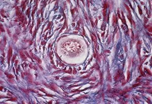
Background imageLight Micrograph Collection: Primordial egg follicle in an ovary

Primordial egg follicle in an ovary
Light micrograph of a primordial follicle (at centre) surrounded by flattened stroma cells in the human ovary. This follicle will form into an oocyte which becomes an egg

Background imageLight Micrograph Collection: Lymphoblastic sarcoma, light micrograph

Lymphoblastic sarcoma, light micrograph
Lymphoblastic sarcoma. Light micrograph of blood cells from bone marrow in a case of lymphoblastic sarcoma. These small lymphoblasts (dark red) characteristically have conspicuous nucleoli

Background imageLight Micrograph Collection: Plasmocyte blood cell, light micrograph

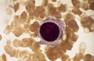
Plasmocyte blood cell, light micrograph. Plasmocytes, also called plasma cells, are derived from stimulated B lymphocytes (white blood cells)

Background imageLight Micrograph Collection: Xanthidium desmids, light micrograph

Xanthidium desmids, light micrograph
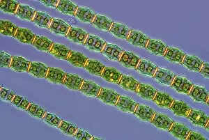
Xanthidium desmids. Polarised light micrograph of a Xanthidium sp. desmid green algae. Desmids are a common group of freshwater single-celled algae that have intricate cell walls

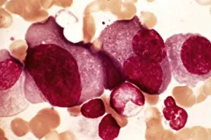
Background imageLight Micrograph Collection: Multiple myeloma, light micrograph

Multiple myeloma, light micrograph
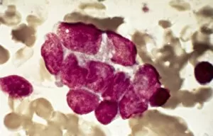
Multiple myeloma. Light micrograph of dystrophy (abnormality) of plasmocyte blood cells from bone marrow in a case of multiple myeloma, also known as Kahlers disease or plasma cell myeloma

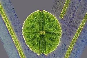
Background imageLight Micrograph Collection: Desmids, light micrograph

Desmids, light micrograph
Desmids. Phase contrast light micrograph of Micrasterias sp. and Hyalotheca sp. desmid green algae. Desmids are a common group of freshwater single-celled algae that have intricate cell walls

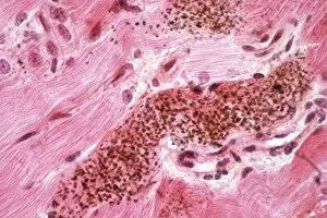
Background imageLight Micrograph Collection: Cushings syndrome, light micrograph

Cushings syndrome, light micrograph
Cushings syndrome. Light micrograph of a section through muscle affected by Cushings syndrome, a hormonal condition caused by overproduction of adrenal gland hormones

Background imageLight Micrograph Collection: Lymphoplasmacytic lymphoma, micrograph

Lymphoplasmacytic lymphoma, micrograph
Lymphoplasmacytic lymphoma. Light micrograph of a mixture of small lymphocytes and plasma cells from bone marrow in a case of lymphoplasmacytic lymphoma, also known as Waldenstroms macroglobulinemia

Background imageLight Micrograph Collection: Ovarian follicle tissue, light micrograph

Ovarian follicle tissue, light micrograph
Ovarian follicle tissue. Light micrograph of a section through tissue from an ovarian follicle, showing a boundary between two layers

Background imageLight Micrograph Collection: Hairy cell leukaemia, light micrograph

Hairy cell leukaemia, light micrograph
Hairy cell leukaemia. Light micrograph of blood cells in a case of hairy cell leukaemia. This sample, treated with May-Grunwald-Giemsa (MGG) stain

Background imageLight Micrograph Collection: Mast cells, light micrograph

Mast cells, light micrograph
Mast cells. Light micrograph of several mast cells (pink rounded objects). These leucocytes (white blood cells) contain the chemical mediators histamine, serotonin and heparin

Background imageLight Micrograph Collection: Chebarkul meteorite fragment C015 / 2862

Chebarkul meteorite fragment C015 / 2862
Chebarkul meteorite fragment seen through a microscope. This research is being carried out at the Vernadsky Institute, Moscow, Russia

Background imageLight Micrograph Collection: Microalgae, light micrograph

Microalgae, light micrograph
Microalgae. Differential interference contrast (DIC) micrograph of Botryococcus braunii (round), and other species of green alga. Magnification: x278, when printed 10 centimetres wide

Background imageLight Micrograph Collection: Cervix, light micrograph

Cervix, light micrograph
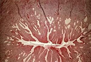
Cervix. Light micrograph of a section through the mucosa of the cervix, the neck of the uterus. This is the endocervix, which borders the uterus. It is lined with columnar epithelial cells

Background imageLight Micrograph Collection: Acute erythroid leukaemia, micrograph

Acute erythroid leukaemia, micrograph
Acute erythroid leukaemia. Light micrograph of blood cells from bone marrow in a case of acute erythroid leukaemia. The cells include dystrophic (degenerated) erythroblasts (nuclei stained dark red)

Background imageLight Micrograph Collection: Closterium desmid, light micrograph

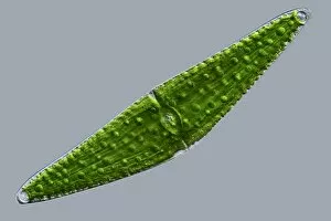
Closterium desmid, light micrograph
Closterium desmid. Differential interference contrast (DIC) micrograph of a Closterium sp. desmid green alga. Desmids are a common group of freshwater single-celled algae that have intricate cell

Background imageLight Micrograph Collection: Acute lymphoblastic leukaemia, micrograph

Acute lymphoblastic leukaemia, micrograph
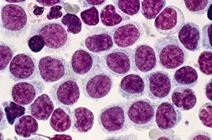
Acute lymphoblastic leukaemia. Light micrograph of blood cells from bone marrow in a case of acute lymphoblastic leukaemia. Leukaemia is a cancer where certain blood cells form in excess

Background imageLight Micrograph Collection: Malignant histiocytosis, light micrograph

Malignant histiocytosis, light micrograph
Malignant histiocytosis. Light micrograph of blood cells in a case of malignant histiocytosis. Hystiocytosis is an excess number of histiocytes

Background imageLight Micrograph Collection: Haematococcus algae, light micrograph

Haematococcus algae, light micrograph
Haematococcus algae. Differential interference contrast (DIC) micrograph of a colony of Haematococcus sp. green alga. Magnification: x220, when printed 10 centimetres wide

Background imageLight Micrograph Collection: Blood cells, light micrograph

Blood cells, light micrograph
Blood cells. Light micrograph of red blood cells (erythrocytes, light blue) and white blood cells (leucocytes, nuclei stained purple)

Background imageLight Micrograph Collection: Chediak-Higashi syndrome, micrograph

Chediak-Higashi syndrome, micrograph
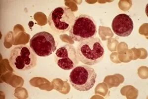
Chediak-Higashi syndrome. Light micrograph of blood cells in a case of Chediak-Higashi syndrome. The cells include neutrophil white blood cells with abnormal granules

Background imageLight Micrograph Collection: Metastatic breast cancer, micrograph

Metastatic breast cancer, micrograph
Metastatic breast cancer. Light micrograph of blood cells in a case of metastatic (secondary) medullary breast cancer. Medullary breast cancer is a rare form of breast cancer that is diagnosed by

Background imageLight Micrograph Collection: Ciliate protozoan, light micrograph

Ciliate protozoan, light micrograph
Ciliate protozoan. Darkfield illuminated light micrograph of a ciliate protozoan, showing green algae (green) that it has ingested

Background imageLight Micrograph Collection: Metastatic lung cancer, micrograph

Metastatic lung cancer, micrograph
Metastatic lung cancer. Light micrograph of blood cells in a case of metastatic (secondary) medullary lung cancer. Medullary cancer (carcinoma)

Background imageLight Micrograph Collection: Multiple sclerosis, light micrograph

Multiple sclerosis, light micrograph
Multiple sclerosis. Light micrograph of a section through the brain showing a large area of demyelinated nerve fibres (light pink, top) due to multiple sclerosis

Background imageLight Micrograph Collection: Pineal gland, light micrograph

Pineal gland, light micrograph. The pineal gland is a small endocrine gland located near to the centre of the brain, between the two hemispheres

Background imageLight Micrograph Collection: Alders anomaly, light micrograph

Alders anomaly, light micrograph
Alders anomaly. Light micrograph of an abnormal lymphocyte white blood cell (centre) from a blood sample in a case of Alders anomaly

Background imageLight Micrograph Collection: Bambusina desmids, light micrograph

Bambusina desmids, light micrograph
Bambusina desmids. Polarised light micrograph of a colony of Bambusina sp. desmid green algae. Desmids are a common group of freshwater single-celled algae that have intricate cell walls

Background imageLight Micrograph Collection: Midge eggs, light micrograph

Midge eggs, light micrograph
Midge eggs. Differential interference contrast (DIC) micrograph of the eggs of a non-biting midge (family Chironomidae). Midge refers to many kinds of very small two-winged flies found world-wide

Background imageLight Micrograph Collection: Acute leukaemia, light micrograph

Acute leukaemia, light micrograph
Acute leukaemia. Light micrograph of blood cells from bone marrow in a case of acute leukaemia. These cells include a polynuclear degranulated basophil and myeloblasts

Background imageLight Micrograph Collection: Chebarkul meteorite fragment C015 / 2860

Chebarkul meteorite fragment C015 / 2860
Chebarkul meteorite fragment seen through a microscope. This research is being carried out at the Vernadsky Institute, Moscow, Russia

Background imageLight Micrograph Collection: Water droplets, light micrograph

Water droplets, light micrograph. Differential interference contrast (DIC) micrograph of water droplets on a surface. Magnification: x278, when printed 10 centimetres wide

Background imageLight Micrograph Collection: Thyroid follicles, light micrograph

Thyroid follicles, light micrograph
Thyroid follicles. Light micrograph of a section through a thyroid gland showing the follicles. The follicles are lined by a single layer of cuboidal epithelial cells

Background imageLight Micrograph Collection: Chronic myeloid leukaemia, micrograph

Chronic myeloid leukaemia, micrograph
Chronic myeloid leukaemia. Light micrograph of blood cells from bone marrow in a case of chronic myeloid leukaemia. Leukaemia is a cancer where certain blood cells form in excess

Background imageLight Micrograph Collection: Granulocyte blood cells, light micrograph

Granulocyte blood cells, light micrograph
Granulocyte blood cells. Light micrograph of two types of granulocytes, a type of white blood cell (leucocyte). At left is a neutrophil, with an eosinophil at right

Background imageLight Micrograph Collection: Reticulosarcoma, light micrograph

Reticulosarcoma, light micrograph
Reticulosarcoma. Light micrograph of reticulocyte blood cells (red, one at upper left) from a lymphatic ganglion in a case of reticulosarcoma

Background imageLight Micrograph Collection: Langerhans cell histiocytosis, micrograph

Langerhans cell histiocytosis, micrograph
Langerhans cell histiocytosis. Light micrograph of blood cells in a case of Langerhans cell histiocytosis (LCH), also called X-type histiocytosis

Background imageLight Micrograph Collection: Rotifer, light micrograph

Rotifer, light micrograph
Rotifer. Darkfield illuminated light micrograph of a rotifer (phylum Rotifera). Rotifers (phylum Rotifera) are microscopic aquatic animals that are related to roundworms

Background imageLight Micrograph Collection: Hodgkins disease, light micrograph

Hodgkins disease, light micrograph
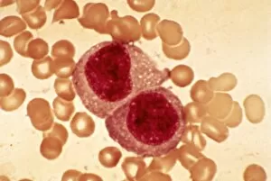
Hodgkins disease. Light micrograph of two non-dystrophic histiocyte blood cells (centre) in a case of Hodgkins disease, also called Hodgkins lymphoma

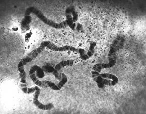
Background imageLight Micrograph Collection: Chromosomes, light micrograph C016 / 6354

Chromosomes, light micrograph C016 / 6354
Chromosomes, light micrograph. This micrograph was obtained in 1934 during studies of chromosome structure in the salivary glands of Sciara (a genus of fungus gnats)

Background imageLight Micrograph Collection: Gastritis, light micrograph C016 / 0508

Gastritis, light micrograph C016 / 0508
Gastritis. Light micrograph of a section through an inflamed human stomach (gastritis), showing an abnormal accumulation of white blood cells (leucocytes)

Background imageLight Micrograph Collection: Corpus luteum of ovary, light micrograph C016 / 0527

Corpus luteum of ovary, light micrograph C016 / 0527
Corpus luteum of ovary. Light micrograph of a section through steroid-secreting luteal cells in the corpus luteum of an ovary. The corpus luteum develops from an ovulated follicle


All Professionally Made to Order for Quick Shipping

Capturing the intricate beauty of biological structures, a light micrograph reveals the mesmerizing complexity of cerebellum tissue. Delicate nerve and glial cells intertwine like a finely woven tapestry, forming the foundation for neurological function. In another stunning image, copper and magnesium sulphate crystals shimmer under the lens of a light microscope, showcasing their crystalline elegance. Moving beyond human anatomy, an awe-inspiring glimpse into early development is unveiled through a light micrograph of a human blastocyst. This embryonic stage brims with promise and potential as it prepares to embark on its journey towards life. Immunofluorescent LM unveils vibrant hues that illuminate neurons and astrocytes in breathtaking detail. These vital components of our nervous system come alive under fluorescent markers, revealing their interconnectedness in supporting brain function. Stepping away from biology but not lacking in fascination, caffeine crystals take center stage in yet another captivating light micrograph. Their jagged edges and distinct patterns mirror the stimulating effects they have on our bodies. Venturing deeper into brain tissue exploration, hippocampus tissue emerges as an enchanting subject for study. Its convoluted structure houses memories and emotions while providing insight into cognitive processes that shape who we are. Glial cells take on an ethereal quality when observed through confocal light microscopy. The interplay between these supportive cells becomes apparent as they weave together like delicate threads within neural networks. HeLa cells become protagonists under the gaze of a light microscope C017/8299 - immortalized cell lines that have revolutionized medical research since their discovery over half a century ago. Their unique characteristics continue to unlock mysteries about cancer and other diseases plaguing humanity. Intricacy extends beyond living organisms; even plant stems hold secrets waiting to be revealed by science's lens. A dicotyledon stem showcases its vascular bundles with precision while hinting at nature's ingenious design principles.