
Lignin Collection
"Lignin: The Mighty Support System in Nature's Architecture From the intricate silver birch twig to the robust xylem tissue


All Professionally Made to Order for Quick Shipping
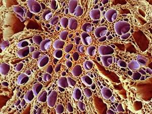
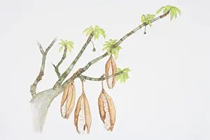
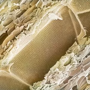
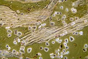
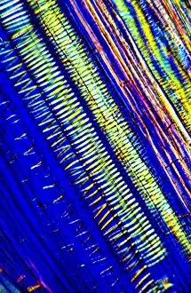
"Lignin: The Mighty Support System in Nature's Architecture From the intricate silver birch twig to the robust xylem tissue, lignin plays a crucial role in providing strength and structure. Under the scanning electron microscope (SEM), we can witness its remarkable presence. In the SEM image of xylem tissue, lignin forms a network of interconnected fibers that transport water and nutrients throughout plants. This complex system ensures their survival and growth. The light micrograph of wood showcases lignin as it gives wood its characteristic hardness and durability. Its presence is vital for trees to withstand external pressures and environmental challenges. Observe the pods hanging from branches of Ceiba pentandra, also known as Kapok Tree – they owe their resilience to lignin. These pods protect seeds until they are ready for dispersal, showcasing nature's ingenious design. Even delicate stems like those found in buttercup flowers possess lignin under scrutiny by SEM. It reinforces their structure, allowing them to stand tall amidst gentle breezes or heavy raindrops. Delving deeper into balsa wood's structure through SEM reveals how lignin contributes to its lightweight yet sturdy composition. This unique combination makes balsa wood an ideal material for various applications such as model making or insulation. Xylem plant cells captured under SEM highlight how lignin provides support within these microscopic structures. It enables efficient water transportation while maintaining stability against internal pressure fluctuations. Elm wood exhibits an abundance when examined closely with SEM – a testament to its exceptional strength and resistance against decay or damage over time. Nature has truly fortified this species with this remarkable substance. As we explore more images of elm wood under SEM, we witness the intricate patterns formed by intertwined layers of cellulose reinforced by abundant deposits of resilient lignin fibers. " Lignin stands out as one of nature's unsung heroes - silently but powerfully contributing to the structural integrity and longevity across various plant species.