
Lining Collection (page 8)
"Lining the Past: From Coats & Fox Terriers to Champagne Hoards" Step back in time to 1927, where coats were lined with fox terrier fur for ultimate warmth and luxury


All Professionally Made to Order for Quick Shipping
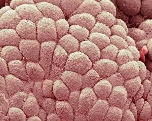
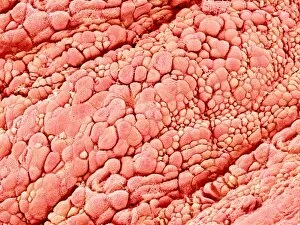
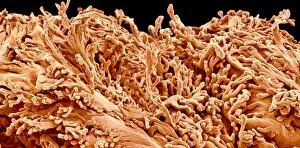
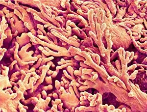
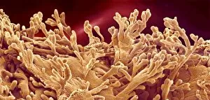
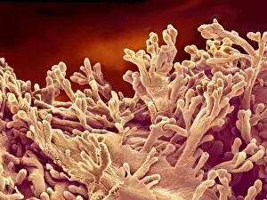
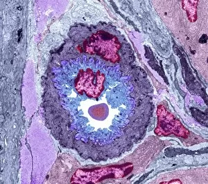
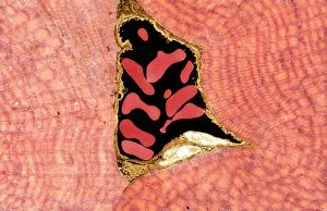
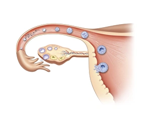
"Lining the Past: From Coats & Fox Terriers to Champagne Hoards" Step back in time to 1927, where coats were lined with fox terrier fur for ultimate warmth and luxury. Meanwhile, hidden treasures of champagne were discovered, forming a hoard that would make any connoisseur's heart skip a beat. Stroll along the picturesque Bouquinistes on Paris Quais, where book and print sellers line the streets, offering literary gems from around the world. Among them lies Lucy Locket's lost pocket - an intriguing mystery waiting to be unraveled. Delve into the microscopic world as we explore the intricate lining of a uterus during menstruation through SEM imagery. Witness nature's remarkable process unfold before your eyes. Admire the grace and elegance captured in a portrait of a geisha holding an ornate umbrella. Every detail meticulously painted, down to each delicate fold lining her kimono. Immerse yourself in history as you step into Plug Street Wood by Matania. Feel the solemnity as soldiers line up amidst towering trees during World War I - their bravery etched forever within these haunting brushstrokes. Experience beauty at its finest with hollyhocks lining a quaint street. Their vibrant colors create an enchanting pathway that beckons you towards hidden wonders just beyond reach. Travel back to West India Docks in 1810, where bustling activity fills every corner. Ships docked side by side while workers diligently load and unload cargo along perfectly aligned lines. Witness sophistication personified as a man peers through his quizzing glass - an accessory once used for discerning details with utmost precision. His sharp gaze reveals secrets only he can uncover. Support local scouts selling Coronation programs; their enthusiasm contagious as they line up eagerly awaiting customers who wish to commemorate this historic event together. Lastly, marvel at our body's resilience as we delve into intestinal linings - protecting us day after day, silently working to keep us healthy and nourished.