
Lower Jaw Collection
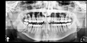
"The Lower Jaw: A Window into Dental Health and Evolutionary History" Panoramic dental X-ray reveals the intricate structure of the lower jaw


All Professionally Made to Order for Quick Shipping
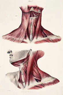
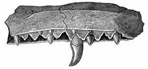
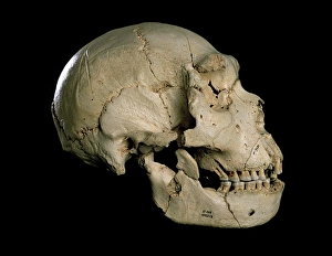
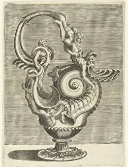
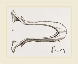
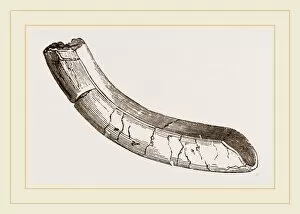
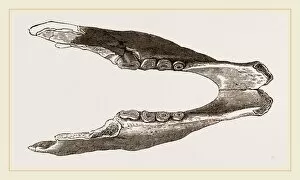
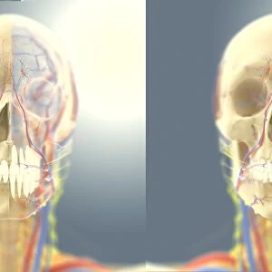
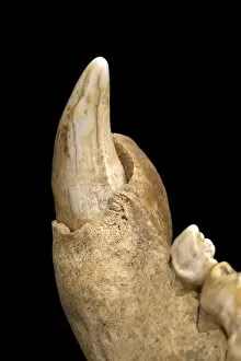
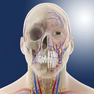
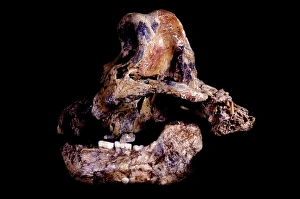
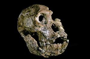
"The Lower Jaw: A Window into Dental Health and Evolutionary History" Panoramic dental X-ray reveals the intricate structure of the lower jaw, providing valuable insights into oral health. The muscles of the neck work in harmony with the lower jaw, facilitating chewing and speaking functions. An artwork depicting a Megalosaurus jaw from the 19th century showcases the impressive size and strength of ancient reptilian jaws. Detailed head and neck anatomy artwork highlights the crucial role played by the lower jaw in supporting facial structures. The skeleton of an Eagle, as depicted in an engraving by Milne-Edwards, demonstrates how different species have adapted their lower jaws for specific purposes. Engravings showcasing human skeletons emphasize that every individual's unique lower jaw contributes to overall skeletal balance and stability. The Homo heidelbergensis skull (Cranium 5) C015 / 6921 provides evidence of early hominid evolution through changes in craniofacial features including the lower jaw. Pages from "The Pictorial Museum of Animated Nature" depict various animal species' lower jaws, highlighting their diversity across nature's kingdom. A broken jaw X-ray C017 / 7557 serves as a reminder of how vulnerable our they can be to injuries requiring specialized care for healing properly. Child's molars and tongue are essential components within a developing mouth - all supported by a growing lower jaw. In Paul Cézanne's painting "Young Man Skull Jeune homme tête de mort, " even art captures fascination with this vital bone structure that has shaped our understanding of dental health and evolutionary history throughout time.